中国组织工程研究 ›› 2018, Vol. 22 ›› Issue (36): 5828-5832.doi: 10.3969/j.issn.2095-4344.0618
• 组织构建实验造模 experimental modeling in tissue construction • 上一篇 下一篇
闭合间断骨钻孔后徒手暴力旋前-外旋型三踝骨折尸体造模
邱 鹏1,成永忠1,刘广伟2,贺 达1,祝建飞1,程 灏1,温建民1,吴丽艳1
- (1中国中医科学院望京医院,北京市 100102;2中医正骨技术北京市重点实验室,北京市 100102)
Establishment of the model of pronation-external rotation trimalleolar fracture after closed osteotomy
Qiu Peng1, Cheng Yongzhong1, Liu Guangwei2, He Da1, Zhu Jianfei1, Cheng Hao1, Wen Jianmin1, Wu Liyan1
- (1Wangjing Hospital of China Academy of Chinese Medical Sciences, Beijing 100102, China; 2Beijing Key Laboratory of Chinese Manipulative Technique, Beijing 100102, China)
摘要:
文章快速阅读:
.jpg) 文题释义:
旋前-外旋三踝骨折:旋前-外旋型三踝骨折主要特点是腓骨骨折水平高于下胫腓联合水平,常复合内踝撕脱骨折或三角韧带断裂、下胫腓分离、后踝骨折。损伤机制是当足位于旋前位时距骨外旋,外侧韧带复合体松弛,首先损伤内踝,可表现为内踝骨折或者三角韧带断裂,暴力继续作用,距骨的内侧壁便可以向前移位,距骨外旋,迫使腓骨沿其纵轴旋转扭曲,腓骨逃离向后外,下胫腓联合及骨间膜从远端向近端撕裂,直致外侧韧带复合体紧张,腓骨无法逃离才发生骨折,暴力继续,造成后踝撕脱骨折或下胫腓后韧带断裂为Ⅳ度。
微创连孔截骨导向器:由日本得岛大学安井夏生、中国骨外固定技术研究所夏和桃医生,几乎在同一个时期发明应用。管状骨电钻打孔微创连孔截骨器,2个孔固定连接在一起,彼此相通,只要使用跟孔径大小一致的截骨针和钻头,连续钻2个孔,拔除另一个克氏针套筒旋转180°,开始下一个骨钻孔,如此循环,可以实现闭合微创截骨,保留软组织的完整性。
文题释义:
旋前-外旋三踝骨折:旋前-外旋型三踝骨折主要特点是腓骨骨折水平高于下胫腓联合水平,常复合内踝撕脱骨折或三角韧带断裂、下胫腓分离、后踝骨折。损伤机制是当足位于旋前位时距骨外旋,外侧韧带复合体松弛,首先损伤内踝,可表现为内踝骨折或者三角韧带断裂,暴力继续作用,距骨的内侧壁便可以向前移位,距骨外旋,迫使腓骨沿其纵轴旋转扭曲,腓骨逃离向后外,下胫腓联合及骨间膜从远端向近端撕裂,直致外侧韧带复合体紧张,腓骨无法逃离才发生骨折,暴力继续,造成后踝撕脱骨折或下胫腓后韧带断裂为Ⅳ度。
微创连孔截骨导向器:由日本得岛大学安井夏生、中国骨外固定技术研究所夏和桃医生,几乎在同一个时期发明应用。管状骨电钻打孔微创连孔截骨器,2个孔固定连接在一起,彼此相通,只要使用跟孔径大小一致的截骨针和钻头,连续钻2个孔,拔除另一个克氏针套筒旋转180°,开始下一个骨钻孔,如此循环,可以实现闭合微创截骨,保留软组织的完整性。摘要
背景:闭合间断骨钻孔截骨方法在临床运用较多,目前却缺少该造模方法的基础实验研究。
目的:建立一种旋前-外旋型三踝骨折模型造模新方法,为闭合截骨造模提供一种新思路、新方法。
方法:对16例典型的旋前-外旋型三踝骨折X射线片骨折线测量来确定钻孔位置,借助微创截骨导向器沿着骨折线在体表投影进行闭合间断骨钻孔后根据其损伤机制徒手暴力造模,同时测量造模过程中的力学参数,通过X射线摄片对模型进行验证。
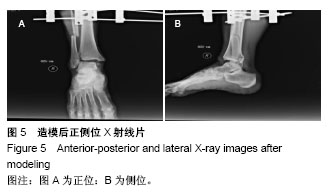
结果与结论:①16个踝关节尸体标本骨折线符合旋前-外旋型三踝骨折,外踝骨折成功率100%,内踝骨折成功率100%,后踝骨折成功率43.75%,下胫腓分离成功率62.50%;②骨折脱位8例,脱位率50%,其中外后脱位2例,外后脱位率12.5%,外脱位6例,外脱位率37.5%;③结果表明,该造模方法成功率高,具有很好的可重复性和可操作性,同时能很好的保留肌肉、肌腱、皮肤等软组织的完整,为三踝骨折手法复位及生物力学研究提供软组织完整的骨折模型,也能为制作闭合骨折模型提供一种新思路、新方法,供他人借鉴参考。
中国组织工程研究杂志出版内容重点:组织构建;骨细胞;软骨细胞;细胞培养;成纤维细胞;血管内皮细胞;骨质疏松;组织工程
ORCID: 0000-0002-6666-5940(邱鹏)
中图分类号:
.jpg)
.jpg)
.jpg)
.jpg)
.jpg) 文题释义:
旋前-外旋三踝骨折:旋前-外旋型三踝骨折主要特点是腓骨骨折水平高于下胫腓联合水平,常复合内踝撕脱骨折或三角韧带断裂、下胫腓分离、后踝骨折。损伤机制是当足位于旋前位时距骨外旋,外侧韧带复合体松弛,首先损伤内踝,可表现为内踝骨折或者三角韧带断裂,暴力继续作用,距骨的内侧壁便可以向前移位,距骨外旋,迫使腓骨沿其纵轴旋转扭曲,腓骨逃离向后外,下胫腓联合及骨间膜从远端向近端撕裂,直致外侧韧带复合体紧张,腓骨无法逃离才发生骨折,暴力继续,造成后踝撕脱骨折或下胫腓后韧带断裂为Ⅳ度。
微创连孔截骨导向器:由日本得岛大学安井夏生、中国骨外固定技术研究所夏和桃医生,几乎在同一个时期发明应用。管状骨电钻打孔微创连孔截骨器,2个孔固定连接在一起,彼此相通,只要使用跟孔径大小一致的截骨针和钻头,连续钻2个孔,拔除另一个克氏针套筒旋转180°,开始下一个骨钻孔,如此循环,可以实现闭合微创截骨,保留软组织的完整性。
文题释义:
旋前-外旋三踝骨折:旋前-外旋型三踝骨折主要特点是腓骨骨折水平高于下胫腓联合水平,常复合内踝撕脱骨折或三角韧带断裂、下胫腓分离、后踝骨折。损伤机制是当足位于旋前位时距骨外旋,外侧韧带复合体松弛,首先损伤内踝,可表现为内踝骨折或者三角韧带断裂,暴力继续作用,距骨的内侧壁便可以向前移位,距骨外旋,迫使腓骨沿其纵轴旋转扭曲,腓骨逃离向后外,下胫腓联合及骨间膜从远端向近端撕裂,直致外侧韧带复合体紧张,腓骨无法逃离才发生骨折,暴力继续,造成后踝撕脱骨折或下胫腓后韧带断裂为Ⅳ度。
微创连孔截骨导向器:由日本得岛大学安井夏生、中国骨外固定技术研究所夏和桃医生,几乎在同一个时期发明应用。管状骨电钻打孔微创连孔截骨器,2个孔固定连接在一起,彼此相通,只要使用跟孔径大小一致的截骨针和钻头,连续钻2个孔,拔除另一个克氏针套筒旋转180°,开始下一个骨钻孔,如此循环,可以实现闭合微创截骨,保留软组织的完整性。