中国组织工程研究 ›› 2019, Vol. 23 ›› Issue (7): 996-1000.doi: 10.3969/j.issn.2095-4344.0576
• 骨组织构建 bone tissue construction • 上一篇 下一篇
幼兔颅盖骨成骨细胞的分离培养及鉴定
艾力麦尔旦•艾尼瓦尔1,李 鹏1,刁兆峰1,木合塔尔•霍加2
- (1新疆维吾尔自治区人民医院口腔科,新疆维吾尔自治区乌鲁木齐市 830001;2深圳市罗湖区人民医院,广东省深圳市 518001)
Isolation, culture and identification of osteoblasts from neonatal rabbit calvarium
Ailimaierdan•Ainiwaer1, Li Peng1, Diao Zhaofeng1, Muhetaer•Huojia2
- (1Department of Stomatology, People’s Hospital of Xinjiang Uygur Autonomous Region, Urumqi 830001, Xinjiang Uygur Autonomous Region, China; 2Luohu District People’s Hospital of Shenzhen, Shenzhen 518001, Guangdong Province, China)
摘要:
文章快速阅读:
.jpg) 文题释义:
差速贴壁法:该方法主要是利用多聚赖氨酸,对成纤维细胞黏附力较强,对许旺细胞黏附力较弱原理,而将成纤维细胞黏附到玻片上,将含有非黏附许旺细胞的上清收集、离心、培养。优点:速度快;许旺细胞产量高;不用抗有丝分裂药物抗血清和补体。由于该方法是在分离获得细胞的同时,就去除了成纤维细胞,因此其速度明显快于其他方法,缺点是成纤维细胞含量较高于其他方法。
改良酶消化法:该实验对传统的成骨细胞培养法进行改良,首先分离骨组织时仔细分离表面纤维结缔及血管软组织,并将组织块儿剪成尽可能小的碎片,胰酶消化适当时间以清除对胰酶消化抵抗能力差的纤维结缔组织、松解成骨细胞,再用Ⅰ型胶原酶消化组织块,从而加快成骨细胞从骨碎片表面爬出的速度,提高得到的细胞数量,加上采用差速贴壁法纯化原代细胞从而消除成纤维细胞、破骨细胞、骨生成细胞等混杂细胞,可以有效提高成骨细胞的纯度。
文题释义:
差速贴壁法:该方法主要是利用多聚赖氨酸,对成纤维细胞黏附力较强,对许旺细胞黏附力较弱原理,而将成纤维细胞黏附到玻片上,将含有非黏附许旺细胞的上清收集、离心、培养。优点:速度快;许旺细胞产量高;不用抗有丝分裂药物抗血清和补体。由于该方法是在分离获得细胞的同时,就去除了成纤维细胞,因此其速度明显快于其他方法,缺点是成纤维细胞含量较高于其他方法。
改良酶消化法:该实验对传统的成骨细胞培养法进行改良,首先分离骨组织时仔细分离表面纤维结缔及血管软组织,并将组织块儿剪成尽可能小的碎片,胰酶消化适当时间以清除对胰酶消化抵抗能力差的纤维结缔组织、松解成骨细胞,再用Ⅰ型胶原酶消化组织块,从而加快成骨细胞从骨碎片表面爬出的速度,提高得到的细胞数量,加上采用差速贴壁法纯化原代细胞从而消除成纤维细胞、破骨细胞、骨生成细胞等混杂细胞,可以有效提高成骨细胞的纯度。摘要
背景:成骨细胞的提取分离尚无公认的高效简便的实验方法。
目的:探讨用改良酶消化法体外分离、培养并鉴定新西兰幼兔颅盖骨成骨细胞的实验方法。
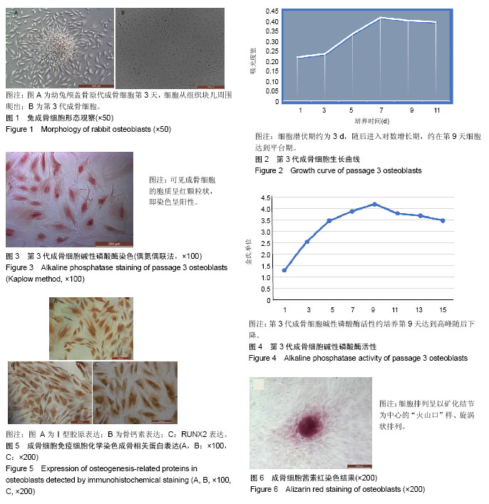
方法:实验利用成纤维细胞胰酶消化时脱壁早,终止消化后贴壁快的特点,采用改良酶消化法分离3 d龄新西兰幼兔颅盖骨成骨细胞并用差速贴壁法纯化培养。倒置显微镜每日观察细胞形态及其生长状况,采用MTT法检测细胞增殖情况并绘制细胞生长曲线,测定碱性磷酸酶含量,免疫组织化学染色法检测Ι型胶原、骨钙素,Runt相关转录因子2(Runt related transcription factor 2,RUNX2)表达,茜素红染色矿化结节等对成骨细胞及其生物化学特性进行鉴定。
结果与结论:①成功分离提取并差速贴壁法纯化培养成骨细胞;②分离培养的成骨细胞贴壁生长,显示正常的成骨细胞形态和生长特征;细胞增殖能力良好;③细胞碱性磷酸酶、Ⅰ型胶原、骨钙素、RUNX2染色均阳性,常规培养21 d茜素红染色形成矿化结节;④结果证实,改良酶消化法培养出的成骨细胞具有典型的成骨细胞特征,成分单一,存活率高。
中国组织工程研究杂志出版内容重点:组织构建;骨细胞;软骨细胞;细胞培养;成纤维细胞;血管内皮细胞;骨质疏松;组织工程
ORCID: 0000-0002-7564-2169(艾力麦尔旦•艾尼瓦尔)
中图分类号:
.jpg) 文题释义:
差速贴壁法:该方法主要是利用多聚赖氨酸,对成纤维细胞黏附力较强,对许旺细胞黏附力较弱原理,而将成纤维细胞黏附到玻片上,将含有非黏附许旺细胞的上清收集、离心、培养。优点:速度快;许旺细胞产量高;不用抗有丝分裂药物抗血清和补体。由于该方法是在分离获得细胞的同时,就去除了成纤维细胞,因此其速度明显快于其他方法,缺点是成纤维细胞含量较高于其他方法。
改良酶消化法:该实验对传统的成骨细胞培养法进行改良,首先分离骨组织时仔细分离表面纤维结缔及血管软组织,并将组织块儿剪成尽可能小的碎片,胰酶消化适当时间以清除对胰酶消化抵抗能力差的纤维结缔组织、松解成骨细胞,再用Ⅰ型胶原酶消化组织块,从而加快成骨细胞从骨碎片表面爬出的速度,提高得到的细胞数量,加上采用差速贴壁法纯化原代细胞从而消除成纤维细胞、破骨细胞、骨生成细胞等混杂细胞,可以有效提高成骨细胞的纯度。
文题释义:
差速贴壁法:该方法主要是利用多聚赖氨酸,对成纤维细胞黏附力较强,对许旺细胞黏附力较弱原理,而将成纤维细胞黏附到玻片上,将含有非黏附许旺细胞的上清收集、离心、培养。优点:速度快;许旺细胞产量高;不用抗有丝分裂药物抗血清和补体。由于该方法是在分离获得细胞的同时,就去除了成纤维细胞,因此其速度明显快于其他方法,缺点是成纤维细胞含量较高于其他方法。
改良酶消化法:该实验对传统的成骨细胞培养法进行改良,首先分离骨组织时仔细分离表面纤维结缔及血管软组织,并将组织块儿剪成尽可能小的碎片,胰酶消化适当时间以清除对胰酶消化抵抗能力差的纤维结缔组织、松解成骨细胞,再用Ⅰ型胶原酶消化组织块,从而加快成骨细胞从骨碎片表面爬出的速度,提高得到的细胞数量,加上采用差速贴壁法纯化原代细胞从而消除成纤维细胞、破骨细胞、骨生成细胞等混杂细胞,可以有效提高成骨细胞的纯度。