中国组织工程研究 ›› 2019, Vol. 23 ›› Issue (7): 958-989.doi: 10.3969/j.issn.2095-4344.1071

• 骨组织构建 bone tissue construction •    下一篇

去卵巢大鼠血清来源外泌体促进原代成骨细胞的增殖

高  坤1,朱文秀2,李  亨1,刘伟东1,李  全1,余伟吉1,王立新1,曹亚飞1   

  1. (1深圳市中医院,广东省深圳市  518000;2北京中医药大学深圳医院,广东省深圳市  518000)
  • 收稿日期:2018-10-07 出版日期:2019-03-08 发布日期:2019-03-08
  • 通讯作者: 曹亚飞,博士,主任医师,深圳市中医院,广东省深圳市 518000
  • 作者简介:高坤,男,1987年生,山东省济南市人,汉族,2016年南方医科大学毕业,博士,医师,主要从事中医骨伤科疾病的临床与基础研究。
  • 基金资助:

    广东省中医药管理局面上项目(20171244),项目负责人:李亨

Exosomes in serum of ovariectomized rats promote primary osteoblast proliferation

Gao Kun1, Zhu Wenxiu2, Li Heng1, Liu Weidong1, Li Quan1, Yu Weiji1, Wang Lixin1, Cao Yafei1   

  1.  (1Shenzhen Traditional Chinese Medicine Hospital, Shenzhen 518000, Guangdong Province, China; 2Shenzhen Hospital of Beijing University of Chinese Medicine, Shenzhen 518000, Guangdong Province, China)
  • Received:2018-10-07 Online:2019-03-08 Published:2019-03-08
  • Contact: Cao Yafei, MD, Chief physician, Shenzhen Traditional Chinese Medicine Hospital, Shenzhen 518000, Guangdong Province, China
  • About author:Gao Kun, MD, Physician, Shenzhen Traditional Chinese Medicine Hospital, Shenzhen 518000, Guangdong Province, China
  • Supported by:

    the General Project of Guangdong Provincial Administration of Traditional Chinese Medicine, No. 20171244 (to LH)

摘要:

文章快速阅读:


文题释义:
外泌体(exosomes):是细胞内部的胞内体与细胞膜融合释放到细胞外,直径大小为30-120 nm的膜性囊泡。
骨重建(Bone remodeling):是指骨组织的形态和密度随着生物力学环境的改变而改变的生理行为。
摘要
背景
:骨代谢过程中,成骨细胞的数量和功能的变化影响骨的生物学特性,外泌体能够通过细胞间的传递进行信号传递,具有促进细胞增殖、分化的潜能。
目的:探讨骨质疏松大鼠血清中外泌体的性质及其对成骨细胞的作用。
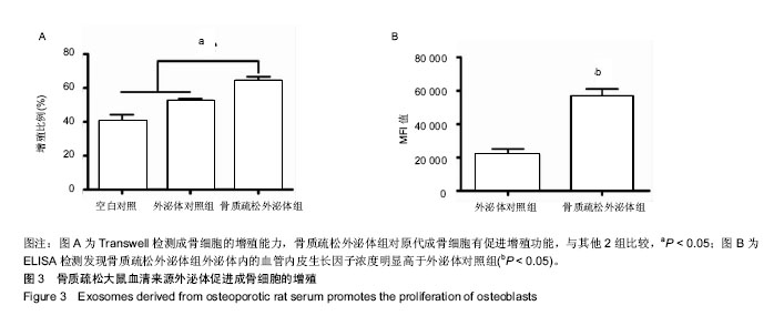
方法:32只成年健康雌性SD大鼠,24 h新生SD大鼠若干只用于原代成骨细胞分离培养,实验动物由广东省医学动物实验中心提供。①建立去卵巢骨质疏松大鼠模型,以假手术组大鼠为对照组。通过ExoQuick提取对照组和骨质疏松大鼠血清中外泌体,用电镜、nanosight、Western blotting进行鉴定;②取对数期的原代成骨细胞接种于96孔细胞培养板中,接种浓度为2×10⁴/孔。实验分为空白对照、外泌体对照组、骨质疏松外泌体组,分别加入DPBS、正常血清外泌体和骨质疏松血清外泌体,100 μL/孔,CO2培养箱培育1 d,采用四甲基偶氮唑盐比色法检测成骨细胞的增殖;酶联免疫吸附试验法检测外泌体中血管内皮生长因子的浓度。
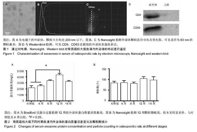
结果与结论:①成功提取大小在100 nm以下外泌体颗粒;②蛋白定量检测12周组外泌体蛋白质量浓度最高;不同时间点外泌体颗粒数12周组略高,但未见明显差异;③骨质疏松大鼠血清来源外泌体与空白对照组及正常外泌体组相比较,明显促进成骨细胞的增殖,且外泌体内血管内皮生长因子升高;④结果说明,骨质疏松大鼠血清中外泌体能够促进成骨细胞的增殖。

中国组织工程研究杂志出版内容重点:组织构建;骨细胞;软骨细胞;细胞培养;成纤维细胞;血管内皮细胞;骨质疏松组织工程
ORCID: 0000-0003-0323-4219(高坤) 

关键词: 骨质疏松, 外泌体, 成骨细胞, 血管内皮生长因子, 组织构建

Abstract:

BACKGROUND: During bone metabolism, changes in the number and function of osteoblasts influence the biological characteristics of bone tissues. Exosomes can mediate cell-to-cell transmission, and have the potential to promote cell proliferation and differentiation.
OBJECTIVE: To study the properties of exosomes in serum of rats with osteoporosis and to investigate their effects on osteoblasts.
METHODS: Several newborn Sprague-Dawley rats at 24 hours of birth were used for primary osteoblast isolation and culture. A rat model of ovariectomized osteoporosis was established in 32 healthy female adult Sprague-Dawley rats. Rats undergoing sham operation were used as control group. All the experimental animals were provided by Guangdong Medical Animal Experimental Center, China. Exosomes were extracted from the serum of control rats and ovariectomized rats using ExoQuick and identified by electron microscopy, nanosight and western blot. Primary osteoblasts in logarithmic phase were seeded into 96-well culture plates with an inoculation concentration of 2×10⁴/well. Then, the cells were divided into blank control group, exosome group, and osteoporosis+exosome group, and DPBS, normal serum-derived exosomes, and osteoporosis serum-derived exosomes, 100 μL/well, were added, respectively. The cells in each group were cultured in a CO2 incubator for 1 day. The proliferation of osteoblasts was detected by MTT colorimetric assay. Enzyme-linked immunosorbent assay was used to detect the concentration of vascular endothelial growth factor in exosomes.
RESULTS AND CONCLUSION: Successfully extracted exosomes particles were below 100 nm in size. The serum exosome protein concentration in osteoporotic rats was highest at 12 weeks after ovariectomized surgery. The number of exosome particles was also highest at 12 weeks after ovariectomized surgery, but there was no significant difference in the osteoporotic rats at different time points. Compared with the blank control and exosome group, exosomes from the serum of osteoporotic rats significantly promoted the proliferation of osteoblasts, and the level of vascular endothelial growth factor in exosomes increased. These findings indicate that exosomes from the serum of osteoporotic rats truly promote the proliferation of osteoblast.

中国组织工程研究杂志出版内容重点:组织构建;骨细胞;软骨细胞;细胞培养;成纤维细胞;血管内皮细胞;骨质疏松组织工程

Key words: Osteoporosis, Exosomes, Osteoblasts, Vascular Endothelial Growth Factors, Tissue Engineering

中图分类号: