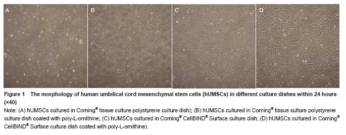
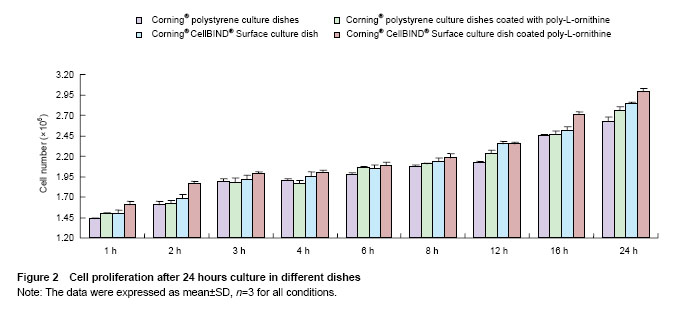
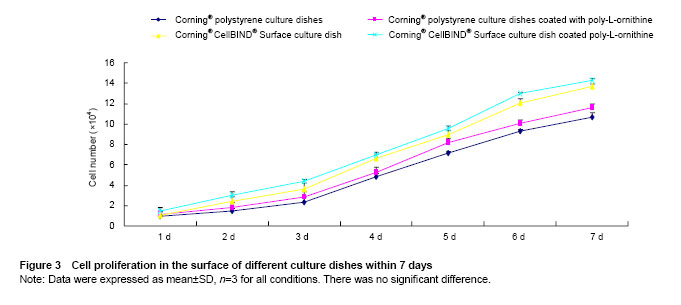
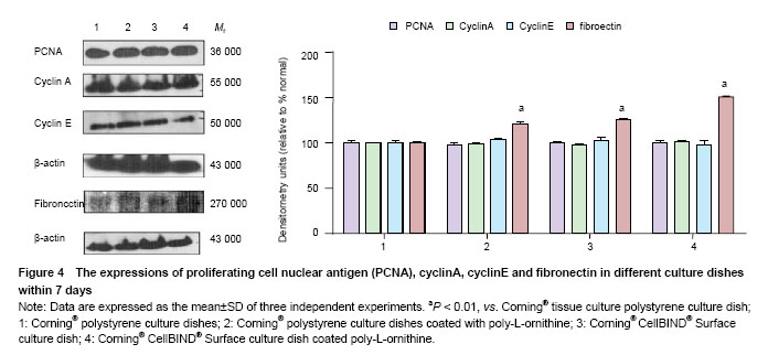
| [1]Ding DC, Shyu WC, Lin SZ. Mesenchymal stem cells. Cell Transplant. 2011;20(1):5-14. [2]Ma L, Feng XY, Cui BL, et al. Human umbilical cord Wharton's Jelly-derived mesenchymal stem cells differentiation into nerve-like cells. Chin Med J (Engl). 2005;118(23):1987-1993.[3]Liu S, Yuan M, Hou K, et al. Immune characterization of mesenchymal stem cells in human umbilical cord Wharton's jelly and derived cartilage cells. Cell Immunol. 2012; 278(1-2):35-44. [4]Song WW, Bai H, Wang CB, et al. Effects of hypoxia on the proliferation of human bone marrow mesenchymal stem cells. Zhonghua Yi Xue Za Zhi. 2010;90(30):2149- 2152.[5]Mitchell KE, Weiss ML, Mitchell BM, et al. Matrix cells from Wharton's jelly form neurons and glia. Stem Cells. 2003; 21(1):50-60.[6]Poulsson AH, Mitchell SA, Davidson MR, et al. Attachment of human primary osteoblast cells to modified polyethylene surfaces. Langmuir. 2009;25(6):3718-3727. [7]Shen M, Horbett TA. The effects of surface chemistry and adsorbed proteins on monocyte/macrophage adhesion to chemically modified polystyrene surfaces. J Biomed Mater Res. 2001;57(3):336-345.[8]Pon-On W, Charoenphandhu N, Teerapornpuntakit J, et al. In vitro study of vancomycin release and osteoblast-like cell growth on structured calcium phosphate-collagen. Mater Sci Eng C Mater Biol Appl. 2013;33(3):1423-1431.[9]Rumian L, Wojak I, Scharnweber D, et al. Resorbable scaffolds modified with collagen type I or hydroxyapatite: in vitro studies on human mesenchymal stem cells. Acta Bioeng Biomech. 2013;15(1):61-67.[10]Gotman I, Ben-David D, Unger RE, et al. Mesenchymal stem cell proliferation and differentiation on load-bearing trabecular Nitinol scaffolds. Acta Biomater. 2013;9(9): 8440-8448. [11]Long X, Matsumoto R, Yang P, et al. Effect of human mesenchymal stem cells on the growth of HepG2 and Hela cells. Cell Struct Funct. 2013;38(1):109-121. [12]Dolley-Sonneville PJ, Romeo LE, Melkoumian ZK. Synthetic surface for expansion of human mesenchymal stem cells in xeno-free, chemically defined culture conditions. PLoS One. 2013;8(8):e70263. [13]Estrada JC, Torres Y, Benguría A, et al. Human mesenchymal stem cell-replicative senescence and oxidative stress are closely linked to aneuploidy. Cell Death Dis. 2013;4:e691. [14]Capra E, Beretta R, Parazzi V, et al. Changes in the proteomic profile of adipose tissue-derived mesenchymal stem cells during passages. Proteome Sci. 2012;10(1):46. [15]Harnett EM, Alderman J, Wood T. The surface energy of various biomaterials coated with adhesion molecules used in cell culture. Colloids Surf B Biointerfaces. 2007;55(1): 90-97.[16]Favi PM, Benson RS, Neilsen NR, et al. Cell proliferation, viability, and in vitro differentiation of equine mesenchymal stem cells seeded on bacterial cellulose hydrogel scaffolds. Mater Sci Eng C Mater Biol Appl. 2013;33(4):1935-1944. [17]Daley WP, Peters SB, Larsen M. Extracellular matrix dynamics in development and regenerative medicine. J Cell Sci. 2008;121(Pt 3):255-264. [18]Page-McCaw A, Ewald AJ, Werb Z. Matrix metalloproteinases and the regulation of tissue remodelling. Nat Rev Mol Cell Biol. 2007;8(3):221-233.[19]van Kooten TG, Spijker HT, Busscher HJ. Plasma-treated polystyrene surfaces: model surfaces for studying cell-biomaterial interactions. Biomaterials. 2004;25(10): 1735-1747.[20]Spoerke ED, Stupp SI. Synthesis of a poly(L-lysine)-calcium phosphate hybrid on titanium surfaces for enhanced bioactivity. Biomaterials. 2005; 26(25):5120-5129.[21]Lu H, Guo L, Kawazoe N, et al. Effects of poly(L-lysine), poly(acrylic acid) and poly(ethylene glycol) on the adhesion, proliferation and chondrogenic differentiation of human mesenchymal stem cells. J Biomater Sci Polym Ed. 2009; 20(5-6):577-589. [22]Steele JG, Dalton BA, Johnson G, et al. Polystyrene chemistry affects vitronectin activity: an explanation for cell attachment to tissue culture polystyrene but not to unmodified polystyrene. J Biomed Mater Res. 1993;27(7): 927-940.[23]Xu Y, Xiao Q, Tian Y, et al. Biological effects of the extracellular matrix on rat bone marrow mesenchymal stem cells. US Chin J Lymphology Oncol. 2007;10(1):26-28.[24]Shadpour H, Sims CE, Thresher RJ, et al. Sorting and expansion of murine embryonic stem cell colonies using micropallet arrays. Cytometry A. 2009;75(2):121-129. [25]Wang Y, Young G, Bachman M, et al. Collection and expansion of single cells and colonies released from a micropallet array. Anal Chem. 2007;79(6):2359-2366. [26]Xu W, Luikart AM, Sims CE, et al. Contact printing of arrayed microstructures. Anal Bioanal Chem. 2010;397(8): 3377-3385.[27]Gunn NM, Bachman M, Li GP, et al. Fabrication and biological evaluation of uniform extracellular matrix coatings on discontinuous photolithography generated micropallet arrays. J Biomed Mater Res A. 2010;95(2):401-412. [28]Shadpour H, Sims CE, Allbritton NL. Enrichment and expansion of cells using antibody-coated micropallet arrays. Cytometry A. 2009;75(7):609-618. [29]Beaulieu I, Geissler M, Mauzeroll J. Oxygen plasma treatment of polystyrene and Zeonor: substrates for adhesion of patterned cells. Langmuir. 2009;25(12): 7169-7176.[30]Kleinhans C, Barz J, Wurster S, et al. Ammonia plasma treatment of polystyrene surfaces enhances proliferation of primary human mesenchymal stem cells and human endothelial cells. Biotechnol J. 2013;8(3):327-337. [31]Lee JH, Jung HW, Kang IK, et al. Cell behaviour on polymer surfaces with different functional groups. Biomaterials. 1994;15(9):705-711.[32]Jankowski RJ, Haluszczak C, Trucco M, et al. Flow cytometric characterization of myogenic cell populations obtained via the preplate technique: potential for rapid isolation of muscle-derived stem cells. Hum Gene Ther. 2001;12(6):619-628.[33]Yun JK, DeFife K, Colton E, et al. Human monocyte/macrophage adhesion and cytokine production on surface-modified poly(tetrafluoroethylene/ hexafluoropropylene) polymers with and without protein preadsorption. J Biomed Mater Res. 1995;29(2):257-268.[34]DeFife KM, Yun JK, Azeez A, et al. Adhesion and cytokine production by monocytes on poly(2-methacryloyloxyethyl phosphorylcholine-co-alkyl methacrylate)-coated polymers. J Biomed Mater Res. 1995;29(4):431-439.[35]Lee JH, Lee HB. A wettability gradient as a tool to study protein adsorption and cell adhesion on polymer surfaces. J Biomater Sci Polym Ed. 1993;4(5):467-481.[36]Fisher RP. The CDK network: linking cycles of cell division and gene expression. Genes Cancer. 2012;3(11-12):731- 738. [37]Opas M, Dziak E. Adhesion, spreading, and proliferation of cells on protein carpets: effects of stability of a carpet. In Vitro Cell Dev Biol. 1991;27A(11):878-885.[38]Jenney CR, Anderson JM. Adsorbed serum proteins responsible for surface dependent human macrophage behavior. J Biomed Mater Res. 2000;49(4):435-447.[39]Flanagan LA, Rebaza LM, Derzic S, et al. Regulation of human neural precursor cells by laminin and integrins. J Neurosci Res. 2006;83(5):845-856. |
.jpg)