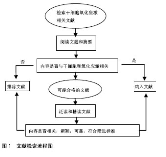
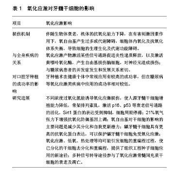
| [1] Collart-Dutilleul PV, Chaubron F, Vos JD, et al. Allogenic banking of dental pulp stem cells for innovative therapeutics. World J Stem Cells. 2015; 7(7): 1010–1021.[2] Rodas-Junco BA, Canul-Chan M, Rojas-Herrera RA, et al. Stem cells from dental pulp: what epigenetics can do with your tooth. Front Physiol. 2017; 8: 999.[3] 张蕾,董小伟,郭凌郧,等. PEP-1-CAT抑制氧化应激条件下大鼠骨髓间充质干细胞的凋亡[J]. 湖北医药学院学报,2012,31(4):285-291.[4] 卢文艺,赵明峰.氧化应激对骨髓间充质干细胞的影响[J].中国医学科学院学报, 2012;34(1):90-94.[5] 肖静雯,王陈飞.N-乙酰氨基葡萄糖转移酶调节O-GlcNAc修饰在牙髓干细胞成骨分化中的作用[J].全科口腔医学杂志:电子版,2016,3(16):11-13.[6] Ratajczak J, Bronckaers A, Dillen Y, et al. The Neurovascular properties of dental stem cells and their importance in dental tissue engineering. Stem Cells Int. 2016; 2016: 9762871.[7] Sonoda S, Yamaza H, Ma L, et al. Interferon-gamma improves impaired dentinogenic and immunosuppressive functions of irreversible pulpitis-derived human dental pulp stem cells. Sci Rep. 2016; 6:19286.[8] Gronthos S, Mankani M, Brahim J, et al. Postnatal human dental pulp stem cells (DPSCs) in vitro and in vivo. Proc Natl Acad Sci. 2000;97:13625-13630.[9] Shi S, Gronthos S. Perivascular niche of postnatal mesenchymal stem cells in human bone marrow and dental pulp. J Bone Miner Res. 2003;18:696-704.[10] 郭红延,吴补领,郭希民,等.大鼠牙髓干细胞的培养和鉴定[J].牙体牙髓牙周病学杂志, 2004,14(5):242-245.[11] 方成志,杨于嘉,姚跃,等.人牙髓干细胞的体外培养及神经样诱导分化[J].中国组织工程研究, 2014,18(23):3723-3726.[12] 李景辉,张方明,张振庭.人恒牙牙髓干细胞分化为脂肪细胞的体外实验研究[J].北京口腔医学,2012,20(3):142-146.[13] D'Aquino R, Graziano A, Sampaolesi M, et al. Human postnatal dental pulp cells co-differentiate into osteoblasts and endotheliocytes: a pivotal synergy leading to adult bone tissue formation. Cell Death Differ.2007;14(6):1162-1171.[14] Otaki S, Ueshima S, Shiraishi K, et al. Mesenchymal progenitor cells in adult human dental pulp and their ability to form bone when transplanted into immunocompromised mice. Cell Biol Int. 2013;31:1191-1197.[15] Tamaoki N, Takahashi K, Tanaka T, et al. Dental pulp cells for induced pluripotent stem cell banking. J Dent Res. 2010;89:773.[16] 何飞,谭颖徽,张纲.人牙髓干细胞的体外培养和鉴定[J].华西口腔医学杂志,2005,23(1):75-78.[17] Zhao Y, Wang L, Jin Y, et al. Fas ligand regulates the immunomodulatory properties of dental pulp stem cells. J Dent Res. 2012;91:948-954.[18] Tatullo M, Marrelli M, Shakesheff KM, et al. Dental pulp stem cells: function, isolation and applications in regenerative medicine. J Tissue Eng Regen Med. 2015;9:1205-1216.[19] Wang J, Wang X, Sun Z, et al. Stem cells from human-exfoliated deciduous teeth can differentiate into dopaminergic neuron-like cells. Stem Cells Dev. 2010;19:1375.[20] Ito K, Yamada Y, Nakamura S, et al. Osteogenic potential of effective bone engineering using dental pulp stem cells, bone marrow stem cells, and periosteal cells for osseointegration of dental implants. Int J Oral Maxillofac Surg. 2011;26:947.[21] 吴中明,木合塔尔•霍加,买布拜木•买买提依明,等.牙髓干细胞在修复牙槽骨缺损的组织学观察[J].石河子大学学报(自科版),2015, 1(33):107-111.[22] Iohara K, Imabayashi K, Ishizaka R, et al. Complete pulp regeneration after pulpectomy by transplantation of CD105+ stem cells with stromal cell-derived factor-1. Tissue Eng Part A. 2011; 17:1911.[23] 王洁,刘加强,房兵.牙髓干细胞在组织工程中的研究进展[J].口腔颌面修复学杂志, 2013,14(3):183-186.[24] 王亦菁,徐莉,金岩,等.体外诱导牙髓干细胞、外胚间充质干细胞向神经分化的研究[J].牙体牙髓牙周病学杂志,2009,19(11):616-618.[25] Leong WK, Henshall TL, Arthur A, et al. Human adult dental pulp stem cells enhance poststroke functional recovery through non-neural replacement mechanisms. Stem Cells Transl Med. 2012;1:177.[26] 孔杰,牛朝诗,杨艳艳,等.骨髓间质干细胞移植对大鼠颅脑损伤后氧化应激的影响[J].立体定向和功能性神经外科杂志,2006,19(3): 149-152.[27] 王全伟,凡文博,王智昊,等.氧化应激与心血管疾病关系的研究进展[J].中国老年学, 2014,34(1):192-194.[28] 邹桂克. bFGF局部应用对2型糖尿病大鼠种植体骨结合的影响[D].第四军医大学,2011.[29] Nevins ML, Karimbux NY, Weber HP, et al. Wound healing around endosseous implants in experimental diabetes. Oral Surg Oral Med Oral Pathol Oral Radiol Endod. 1998;87:293.[30] Mccracken M, Lemons JE, Rahemtulla F, et al. Bone response to titanium alloy implants placed in diabetic rats. Int J Oral Maxillofac Implants. 2000;15:345.[31] Jian Z, Li K, Liu L, et al. Heme oxygenase-1 protects human melanocytes from H2O2-induced oxidative stress via the Nrf2-ARE pathway. J Invest Dermatol. 2011;131:1420.[32] Kim JH, Choi W, Lee JH, et al. Astaxanthin Inhibits H_2O_2-Mediated Apoptotic Cell Death in Mouse Neural Progenitor Cells via Modulation of P38 and MEK Signaling Pathways. J Microbiol Biotechnol. 2009;19:1355-1363.[33] 徐克,冯桂娟,冯兴梅,等.过氧化氢刺激人牙髓干细胞的衰老进程[J].中国组织工程研究, 2016,20(10):1481-1487.[34] Alami M, Viña-Almunia J, Gambini J, et al. Activation of p38, p21, and NRF-2 mediates decreased proliferation of human dental pulp stem cells cultured under 21% O2. Stem Cell Reports. 2014;3:566-573.[35] Jung CY, Yeon LJ, Pyoung CC, et al. Cell-penetrating superoxide dismutase attenuates oxidative stress-induced senescence by regulating the p53-p21Cip1pathway and restores osteoblastic differentiation in human dental pulp stem cells.Int J Nanomedicine. 2012;7:5091-5106.[36] Levine AJ. p53, the cellular gatekeeper for growth and division. Cell. 1997;88:323-331.[37] Yang DG, Liu L, Zheng XY. Cyclin-dependent kinase inhibitor p16 INK4a and telomerase may co-modulate endothelial progenitor cells senescence. Ageing Res Rev. 2008;7:137-146.[38] Mimeault M, Batra SK. Recent insights into the molecular mechanisms involved in aging and the malignant transformation of adult stem/progenitor cells and their therapeutic implications. Ageing Res Rev. 2009;8:94-112.[39] Ma D, Cui L, Gao J, et al. Proteomic analysis of mesenchymal stem cells from normal and deep carious dental pulp. PLoS One. 2014;9:e97026.[40] 凌均棨,彭正军,刘路.细胞重编程及其影响因素[J].国际口腔医学杂志,2014,41(3):300-303.[41] Ond?ej V, Protivánková I, Piterková J, et al. Recondensation level of repetitive sequences in the plant protoplast nucleus is limited by oxidative stress. J Exp Bot.2010;61:2395.[42] Zhang W, Su X, Gao Y, et al. Berberine protects mesenchymal stem cells against hypoxia-induced apoptosis in vitro. Biol Pharm Bull. 2009;32:1335-1342.[43] Wei H, Li Z, Hu S, et al. Apoptosis of mesenchymal stem cells induced by hydrogen peroxide concerns both endoplasmic reticulum stress and mitochondrial death pathway through regulation of caspases, p38 and JNK. J Cell Biochem. 2010;111: 967-978.[44] 黄文秋,黄宏,徐祥,等.mTOR及其下游信号通路在骨髓间充质干细胞氧化应激损伤中的变化及作用[J].第三军医大学学报,2013,35(2): 114-118.[45] 胡明,黎佼,刘宁宁,等.miR-486-5p在氧化应激引起人骨髓间充质干细胞凋亡中的作用[J]. 中国病理生理杂志,2015,31(3):524-529.[46] 束波,刘志江,范芳.基质细胞衍生因子1预处理抑制大鼠骨髓间充质干细胞的凋亡[J].中国组织工程研究,2012,16(32): 5903-5908.[47] Dai Z, Li Y, Quarles LD, et al. Resveratrol enhances proliferation and osteoblastic differentiation in human mesenchymal stem cells via ER-dependent ERK1/2 activation. Phytomedicine. 2007;14: 806-814.[48] Denu RA, Hematti P. Effects of oxidative stress on mesenchymal stem cell biology. Oxid Med Cell Longev.2016;2016:1-9.[49] Volonte D, Zou H, Bartholomew JN, et al. Oxidative stress-induced inhibition of sirt1 by caveolin-1 promotes p53-dependent premature senescence and stimulates the secretion of interleukin 6 (IL-6). J Biol Chem. 2015;290:4202.[50] Kao CL, Chen LK, Chang YL, et al. Resveratrol protects human endothelium from H(2)O(2)-induced oxidative stress and senescence via SirT1 activation. J Thromb Thrombolysis. 2010; 17:970. |
.jpg)
.jpg)