| [1] Sun C,Yu D,Ye W,et al.The short stature homeobox 2 (Shox2)-bone morphogenetic protein (BMP) pathway regulates dorsal mesenchymal protrusion development and its temporary function as a pacemaker during cardiogenesis.J Biol Chem.2015;290 (4): 2007-2023. [2] Zhang KK, Xiang M, Zhou L, et al. Gene network and familial analyses uncover a gene network involving Tbx5/Osr1/Pcsk6 interaction in the second heart field for atrial septation.Hum Mol Genet.2016; 25(6):1140-1151.[3] Kelder TP,Vicente-Steijn R,Harryvan TJ,et al.The sinus venosus myocardium contributes to the atrioventricular canal: potential role during atrioventricular node development? J Cell Mol Med. 2015;19(6):1375-1389.[4] Ammirabile G,Tessari A,Pignataro V,et al.Pitx2 confers left morphological, molecular, and functional identity to the sinus venosus myocardium.Cardio Res.2012;93(2): 291-301. [5] Kim JH,Hwang SE,Rodríguez-Vázquez JF,et al.Upper terminal of the inferior vena cava and development of the heart atriums: a study using human embryos.Anat Cell Biol.2014;47(4):236-243.[6] Anderson RH,Cook AC.The structure and components of the atrial chambers.Europace.2007;9 (suppl 6), vi3-vi9. [7] Lescroart F,Mohun T,Meilhac SM,et al.Lineage tree for the venous pole of the heart: clonal analysis clarifies controversial genealogy based on genetic tracing.Circ Res.2012;111 (10): 1313-1322.[8] Christoffels VM,Mommersteeg MT,Trowe MO,et al.Formation of the venous pole of the heart from an Nkx2-5-negative precursor population requires Tbx18.Circ Res.2006;98(12): 1555-1563.[9] Mommersteeg MT,Dominguez JN,Wiese C,et al.The sinus venosus progenitors separate and diversify from the first and second heart fields early in development.Cardiovasc Res. 2010;87 (1): 92-101.[10] O’Rahilly R,Müller F.Developmental Stages in Human Embryos[M].Washington,DC: Carnegie Institution of Washington Publication.1987.[11] Soufan AT,van den Hoff MJB,Ruijter JM,et al.Reconstruction of the patterns of gene expression in the developing mouse heart reveals an architectural arrangement that facilitates the understanding of atrial malformations and arrhythmias.Circ Res.2004;95(12):1207-1215.[12] Sizarov A,Anderson RH,Christoffels VM,et al. Three-Dimensional and molecular analysis of the venous pole of the developing human heart.Circulation.2010;122(8): 798-807.[13] Witzel HR,Cheedipudi S,Gao R,et al.Isl2b regulates anterior second heart field development in zebrafish.Sci Rep.2017;7: 41043.[14] Yuan XJ,Qi H,Li X,et al.Disruption of spatiotemporal hypoxic signaling causes congenital heart disease in mice.J Clin Invest. 2017;127 (6):2235-2248.[15] van den Berg G,Moorman AFM.Development of the pulmonary vein and the systemic venous sinus: an interactive 3D overview.PLoS One.2011;6(7):e22055.[16] Jongbloed MR,Mahtab EA,Blom NA,et al.Development of the cardiac conduction system and the possible relation to predilection sites of arrhythmogenesis.Scientific World J. 2008;8:239-269.[17] Ye W,Wang J,Song Y,et al.A common Shox2-Nkx2-5 antagonistic mechanism primes the pacemaker cell fate in the pulmonary vein myocardium and sinoatrial node. Development. 2015;142(14): 2521-2532.[18] Gittenberger-de Groot AC, Mahtab EA, Hahurij ND, et al. Nkx2.5-negative myocardium of the posterior heart field and its correlation with podoplanin expression in cells from the developing cardiac pacemaking and conduction system.Anat Rec.2007;290 (1):115-122.[19] Burns T,Yang Y,Hiriart E,et al.The dorsal mesenchymal protrusion and the pathogenesis of atrioventricular septal defects.J Cardiovasc Dev Dis.2016;3:29. [20] Kelder TP,Vicente-Steijn R,Harryvan TJ,et al.The sinus venosus myocardium contributes to the atrioventricular canal: potential role during atrioventricular node development? J Cell Mol Med. 2015;19 (6):1375-1389. |
.jpg) 文题释义:
心背侧间充质突(dorsal mesenchymal protrusion,DMP):是前肠腹侧与心背系膜之间的间充质细胞向心房背侧壁迁移形成的楔状突起。研究显示有的房室隔缺损病例房室管心内膜垫、原发隔形态正常,而心背侧间充质突体积较小,导致心房原发孔不能正常闭合。由此说明在小鼠胚胎、人胚,DMP对心房分隔具有重要作用。
第二生心区(second heart field,SHF):分布于前肠腹侧间充质、心包腔背侧壁的脏壁中胚层等部位,其作用在于参与形成心房大部、右心室和流出道心肌、主、肺动脉壁的部分平滑肌等,其发育异常可导致永存动脉干等先天性心脏畸形的发生。
文题释义:
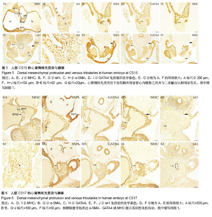
心背侧间充质突(dorsal mesenchymal protrusion,DMP):是前肠腹侧与心背系膜之间的间充质细胞向心房背侧壁迁移形成的楔状突起。研究显示有的房室隔缺损病例房室管心内膜垫、原发隔形态正常,而心背侧间充质突体积较小,导致心房原发孔不能正常闭合。由此说明在小鼠胚胎、人胚,DMP对心房分隔具有重要作用。
第二生心区(second heart field,SHF):分布于前肠腹侧间充质、心包腔背侧壁的脏壁中胚层等部位,其作用在于参与形成心房大部、右心室和流出道心肌、主、肺动脉壁的部分平滑肌等,其发育异常可导致永存动脉干等先天性心脏畸形的发生。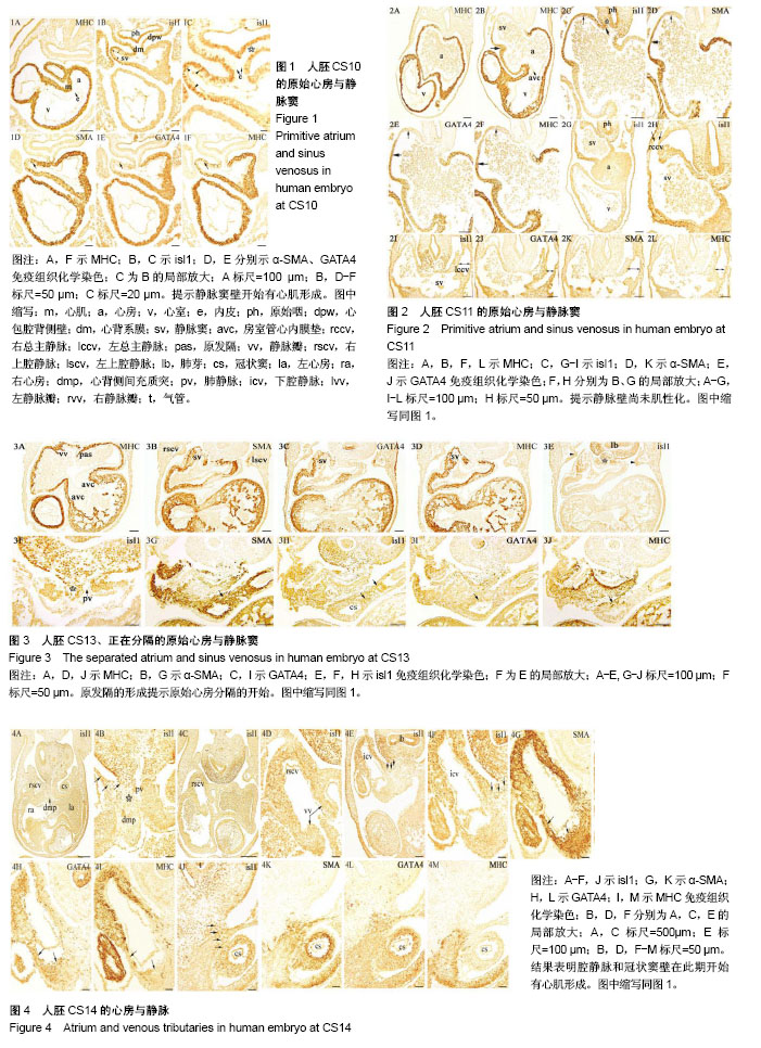
.jpg) 文题释义:
心背侧间充质突(dorsal mesenchymal protrusion,DMP):是前肠腹侧与心背系膜之间的间充质细胞向心房背侧壁迁移形成的楔状突起。研究显示有的房室隔缺损病例房室管心内膜垫、原发隔形态正常,而心背侧间充质突体积较小,导致心房原发孔不能正常闭合。由此说明在小鼠胚胎、人胚,DMP对心房分隔具有重要作用。
第二生心区(second heart field,SHF):分布于前肠腹侧间充质、心包腔背侧壁的脏壁中胚层等部位,其作用在于参与形成心房大部、右心室和流出道心肌、主、肺动脉壁的部分平滑肌等,其发育异常可导致永存动脉干等先天性心脏畸形的发生。
文题释义:
心背侧间充质突(dorsal mesenchymal protrusion,DMP):是前肠腹侧与心背系膜之间的间充质细胞向心房背侧壁迁移形成的楔状突起。研究显示有的房室隔缺损病例房室管心内膜垫、原发隔形态正常,而心背侧间充质突体积较小,导致心房原发孔不能正常闭合。由此说明在小鼠胚胎、人胚,DMP对心房分隔具有重要作用。
第二生心区(second heart field,SHF):分布于前肠腹侧间充质、心包腔背侧壁的脏壁中胚层等部位,其作用在于参与形成心房大部、右心室和流出道心肌、主、肺动脉壁的部分平滑肌等,其发育异常可导致永存动脉干等先天性心脏畸形的发生。