中国组织工程研究 ›› 2021, Vol. 25 ›› Issue (23): 3684-3689.doi: 10.12307/2021.040

• 组织构建实验造模 experimental modeling in tissue construction • 上一篇    下一篇

阳极阻滞电刺激骶神经根重构膀胱功能的作用机制

闫  鹏1,马玉斐2,崔京福2,郝少飞2,刘金辉2,关春雷2,王潇燃3,杨小玉4   

  1. 1郑州大学第五附属医院,河南省郑州市  450000;2郑州市第一人民医院,河南省郑州市  450004;3吉林省人民医院,吉林省长春市  130000;4吉林大学第二医院,吉林省长春市  130041
  • 收稿日期:2019-03-26 修回日期:2019-04-08 接受日期:2020-06-18 出版日期:2021-08-18 发布日期:2021-01-26
  • 通讯作者: 杨小玉,教授,博士生导师,主任医师。吉林大学第二医院,吉林省长春市 130041
  • 作者简介:闫鹏,男,1980年生,吉林省通化市人,汉族,2011年吉林大学毕业,博士,主任医师,主要从事脊髓损伤后膀胱功能重建研究。
  • 基金资助:
    河南省科技厅引智项目(GH2019010),项目负责人:闫鹏

Mechanism of anodic block electrical stimulation of sacral nerve root to reconstruct bladder function

Yan Peng1, Ma Yufei2, Cui Jingfu2, Hao Shaofei2, Liu Jinhui2, Guan Chunlei2, Wang Xiaoran3, Yang Xiaoyu4   

  1. 1The Fifth Affiliated Hospital of Zhengzhou University, Zhengzhou 450000, Henan Province, China; 2Zhengzhou First People's Hospital, Zhengzhou 450004, Henan Province, China; 3Jilin Province People's Hospital, Changchun 130000, Jilin Province, China; 4The Second Hospital of Jilin University, Changchun 130041, Jilin Province, China
  • Received:2019-03-26 Revised:2019-04-08 Accepted:2020-06-18 Online:2021-08-18 Published:2021-01-26
  • Contact: Yang Xiaoyu, Professor, Doctoral supervisor, Chief physician, The Second Hospital of Jilin University, Changchun 130041, Jilin Province, China
  • About author:Yan Peng, MD, Chief physician, The Fifth Affiliated Hospital of Zhengzhou University, Zhengzhou 450000, Henan Province, China
  • Supported by:
    Talent Introduction Project of Henan Provincial Department of Science and Technology, No. GH2019010 (to YP)

摘要:

文题释义:
神经源性膀胱:是指控制排尿功能的中枢神经系统损伤或脊髓损伤后引起的膀胱功能障碍,表现为神经源性逼尿肌过度活动和逼尿肌及括约肌协同运动失调,患者贮尿与排尿功能双重障碍。
阳极阻滞电刺激技术:其原理是用阴极电流刺激神经纤维,将使神经轴索的膜外电位降低,逐渐增大刺激电流,膜电位将超过兴奋的阈电位发生去极化,激发一个动作电位,向远近2个方向上传导。

背景:课题组前期研究表明,阳极阻滞电刺激骶神经根可以改善膀胱储尿和排尿功能,但其具体分子机制未明。
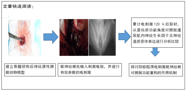
目的:实验采用免疫组织化学染色从蛋白功能角度对膀胱逼尿肌内神经递质受体及神经生长因子表达进行分析,探讨阳极阻滞电刺激骶神经根对膀胱功能重构的作用机制。
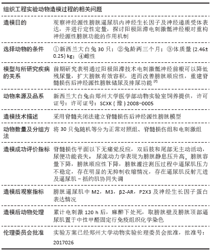
方法:实验将新西兰兔30只随机分为对照组、脊髓损伤组和电刺激组,每组10只,其中脊髓损伤组和电刺激组采用脊髓夹闭法建立脊髓损伤后神经源性膀胱动物模型,电刺激组采用阳极阻滞电刺激骶神经根(300 μs,1.05 mA,20 Hz,ON 5 s,OFF 10 s:间断电刺激,采用开5 s后间歇10 s),累计电刺激骶神经根120 h后取材膀胱逼尿肌,采用免疫组织化学染色从蛋白质功能角度对神经源性膀胱逼尿肌内各种因子进行定性定量分析。
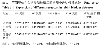
结果与结论:①组织学变化显示,脊髓损伤组取材标本可见膀胱体积明显降低,颜色呈暗褐色,组织表面附着血管网数量下降,膀胱边界模糊,与周围组织部分形成粘连,取材切面可见部分纤维化及组织坏死;对照组与电刺激组形态较为一致,均表现为膀胱体积扩大,色淡红,取材组织表面可见丰富的动静脉血管网,边界清晰,组织内可见较多残留尿液,色微黄且澄清透明,切面与周围组织相比未见异常;②免疫组织化学染色结果显示,M2受体、P2X3受体和神经生长因子受体在脊髓损伤组膀胱逼尿肌中表达明显高于电刺激组和对照组(P < 0.05),M3受体、β2-AR受体在脊髓损伤组膀胱逼尿肌中的表达明显低于电刺激组和对照组(P < 0.05);③结果证实,通过阳极阻滞电刺激骶神经根,可影响神经源性膀胱内神经生长因子及神经递质受体的表达水平,从而影响膀胱逼尿肌的舒缩功能,改善膀胱顺应性,重构脊髓损伤后神经源性膀胱的功能。
https://orcid.org/0000-0002-4251-7847 (闫鹏) 

中国组织工程研究杂志出版内容重点:组织构建;骨细胞;软骨细胞;细胞培养;成纤维细胞;血管内皮细胞;骨质疏松;组织工程

关键词: 脊髓损伤, 阳极阻滞, 电刺激, 骶神经根, 免疫组织化学, 膀胱功能, 重构

Abstract: BACKGROUND: Our previous findings have shown that anodic block electrical stimulation of sacral nerve root can adjust urine storage and urination, but the specific molecular mechanism is unknown.
OBJECTIVE: To analyze the expression of neurotransmitter receptors and nerve growth factor in the bladder detrusor using immunohistochemistry from the perspective of protein function and to explore the mechanism of anodic block electrical stimulation of sacral nerve root on bladder function reconstruction. METHODS: Thirty New Zealand rabbits were randomly divided into control group (n=10), spinal cord injury group (n=10) and electrical stimulation group (n=10). In the latter two groups, animal models of post-spinal cord injury neurogenic bladder were made using spinal cord clippings. In the electrical stimulation group, the sacral nerve root was subjected to an intermittent electrical stimulation (300 μs, 1.05 mA, 20 Hz), 5 seconds on and 10 seconds off for 120 hours. Then, the bladder detrusor was harvested. The expression of nerve growth factor and neurotransmitter receptor in neurogenic bladder detrusor was analyzed qualitatively and quantitatively by immunohistochemistry from the perspective of protein function
RESULTS AND CONCLUSION: In the spinal cord injury group, there was a significantly smaller, dark-brown bladder, with reduced number of vessel plexuses on the tissue surface. The bladder boundary was blurred, with some adhesion to the surrounding tissue, and partial fibrosis and tissue necrosis on the section surface were observed. The morphology of the control group and the electrical stimulation group was relatively consistent, both with enlarged bladder volume and light red in color. Abundant arteriovenous networks were visible on the surface of the tissue. The boundary was clear. Residual urine amount was increased in the tissue. The color was yellowish and transparent. There was no abnormality in the section compared with the surrounding tissues. Immunohistochemical findings shown that the expression of M2 receptor, P2X3 receptor and nerve growth factor receptor in bladder detrusor muscle in the spinal cord injury group was significantly higher than that in the electric stimulation group and control group (P < 0.05), while the expression of M3 receptor and β2-AR receptor in bladder detrusor muscle in spinal cord injury group was significantly lower than that in the electric stimulation group and control group (P < 0.05). To conclude, electrical stimulation of the sacral nerve root by anodic block can adjust the expression of nerve growth factor and neurotransmitters in neurogenic bladder, thereby influencing the systolic and diastolic function of bladder detrusor, improving bladder compliance and remodeling the function of neurogenic bladder after spinal cord injury. 

Key words: spinal cord injury, anodic block, electrical stimulation, sacral nerve root, immunohistochemistry, bladder function, remodeling

中图分类号: