[1] JI WC, JIANG WT, LI M, et al. miR-21 deficiency contributes to the impaired protective effects of obese rat mesenchymal stem cell-derived exosomes against spinal cord injury. Biochimie. 2019;167(6):171-178.
[2] AHUJA CS, WILSON JR, NORI S, et al. Traumatic spinal cord injury. Nat Rev Dis Primers. 2017;3(14):170-180.
[3] 杨俊松,郝定均,刘团江,等.急性脊髓损伤的临床治疗进展[J].中国脊柱脊髓杂志,2018,28(4):368-373.
[4] ANWAR MA, AL STS, EID AH, et al. Inflammogenesis of Secondary Spinal Cord Injury. Front Cell Neurosci. 2016;9(10):98-102.
[5] GORGEY AS, KHALIL RE, LESTER RM, et al. Paradigms of Lower Extremity Electrical Stimulation Training After Spinal Cord Injury. J Vis Exp. 2018; 8(132):1076-1082.
[6] LUNDELL LS, SAVIKJ M, KOSTOVSKI E, et al. Protein translation,proteolysis and autophagy in human skeletal muscle atrophy after spinal cord injury. Acta Physiol (Oxf). 2018;16(7):192-202.
[7] MAO Y, TONKIN RS, NGUYEN T, et al. Systemic Administration of Connexin43 Mimetic Peptide Improves Functional Recovery after Traumatic Spinal Cord Injury in Adult Rats. J Neurotrauma. 2017;34(3):707-719.
[8] YANG Z, ZHANG A, DUAN H, et al. NT 3-chitosan elicits robust endogenous neurogenesis to enable functional recovery after spinal cord injury. Proc Natl Acad Sci U S A. 2015;112(43):13354-13359.
[9] RAO JS, ZHAO C, HANG A, et al. NT 3-chitosan enables de novo regeneration and functional recovery in monkeys after spinal cord injury. Proc Natl Acad Sci U S A. 2018;115(24):E5595-E5604.
[10] DUAN H, GE W, ZHANG A, et al. Transcriptome analyses reveal molecular mechanisms underlying functional recovery after spinal cord injury. Proc Natl Acad Sci U S A. 2015;112(43):13360-13365.
[11] WANG Y, WU W, WU X, et al. Remodeling of lumbar motor circuitry remote to a thoracic spinal cord injury promotes locomotor recovery. Elife. 2018;7:e39016.
[12] YUAN S, SHI Z, CAO F, et al. Epidemiological Features of Spinal Cord Injury in China: A Systematic Review. Front Neurol. 2018;9:683.
[13] PATEL NP, HUANG JH. Hyperbaric oxygen therapy of spinal cord injury. Med Gas Res. 2017;7(2):133-143.
[14] MARTINEZ M,ROSSIGNOL S.A dual spinal cord lesion paradigm to study spinal locomotor plasticity in the cat. Ann N Y Acad Sci. 2013;1279(4):127-134.
[15] YOZBATIRAN N, KESER Z, DAVIS M, et al. Transcranial direct current stimulation (tDCS) of the primary motor cortex and robot-assisted arm training in chronic incomplete cervical spinal cord injury: A proof of concept sham-randomized clinical study. NeuroRehabilitation. 2016;39(3):401-411.
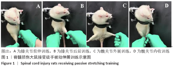
[16] KELLER AV, WAINWRIGHT G, SHUM-SIU A, et al. Disruption of Locomotion in Response to Hindlimb Muscle Stretch at Acute and Chronic Time Points after a Spinal Cord Injury in Rats. J Neurotrauma. 2017;34(3):661-670.
[17] BATTISTUZZO CR, RANK MM, FLYNN JR, et al. Effects Of treadmill training on hindlimb muscles of spinal cord injured mice. Muscle Nerve. 2017; 55(2):232-242.
[18] TINTIGNAC LA, BRENNER HR, RUEGG MA, et al. Mechanisms regulating neuromus- cular junction development and function and causes of muscle wasting. Physiol Rev. 2015;95(3):809-852.
[19] LIU W, CHAKKALAKAL JV. The composition,development and regeneration of neuromuscular junctions. Curr Top Dey Biol. 2018;126(2):99-124.
[20] LARSSON L, DEGENS H, LI M, et al. Sarcopenia: Aging-Related Loss of Muscle Mass and Function. Physiol Rev. 2019;99(1):427-511.
[21] DKS L, SCHOELLER SD, NDS K, et al. Protocol for a scoping review of skin self-care of people with spinal cord injury. BMJ Open. 2017;7(9):e017860.
[22] BOYCE VS, MENDELL LM. Neurotrophic factors in spinal cord injury. Handb Exp Pharmacol. 2014;220(6):443-460.
[23] 范晓华,纪树荣,周红俊,等.减重平板步行训练对完全性脊髓损伤患者下肢骨骼肌萎缩与步行能力的影响[J].中国康复理论与实践,2008, 14(1):50-52.
[24] SOBOTKA S, CHEN J, NYIRENDA T, et al. Outcomes of muscle reinnervation with direct nerve implantation into the native motor zone of the target muscle. J Reconstr Microsurg. 2017;33(2):77-86.
[25] RUDOLF R, DESCHENES MR, SANDRI M,et al. Neuromuscular junction degeneration in muscle wasting. Curr Opin Clin Nutr Metab Care. 2016; 19(3):177-181.
[26] WILSON RJ, DRAKE J C, CUI D, et al. Mitochondrial protein Snitrosation protects against ischaemia reperfusion-induced denervation at neuromuscular junction in skeletal muscle. Free Radic Biol Med. 2018; 117(9):180-190.
[27] MANTHOU M, ABDULLA DS, PAVLOV SP, et al. Whole body vibration (WBV) follo-wing spinal cord injury (SCI) in rats:Timing of intervention. Restor Neurol Neurosci. 2017;35(2):185-216.
[28] 赵博伦.不同起始时间点机器人辅助减重步行训练对脊髓损伤康复作用的研究[D].吉林:吉林大学护理学院,2018.
[29] KELLER AV, WAINWRIGHT G, SHUM-SIU A, et al. Disruption of Locomotion in Response to Hindlimb Muscle Stretch at Acute and Chronic Time Points after a Spinal Cord Injury in Rats. J Neurotrauma. 2017;34(3):661-670.
[30] TOSOLINI AP, MOHANR, MORRIS R. Targeting the full length of the motrend plate regions in the mouse forelimb increases the uptake of Fluoro-Gold into corresponding spinal cord motoneurons. Front Neurol. 2013;4(58):1-10.
[31] NEES TA, TAPPE-THEODOR A, SLIWINSKI C, et al. Early-onset treadmill training reduces mechanical allodynia and modulates calcitonin gene-te- lated peptide fiber density in lamina ll /N in a mouse model of spinal cord contusion injury. Pain. 2016;157(3):687-697.
[32] STUART DG, HULYBORN H. Thomas graham brown (1882-1965),anders lundbery (1920),and the neural control of stepping. Brain Res Rev. 2008; 59(1):74-95.
[33] KIM MK, LEE SA. Underwater treadmill training and gait ability in the normal adult. J Phys Ther Sci. 2017;29(1):67-69.
[34] HACHEM LD, AHUJA CS, FEHLINGS MG, et al. Assessment and management of acute spinal cord injury: From point of injury to rehabilitation. J Spinal Cord Med. 2017;40(6):665-675.
[35] FEHLINGS MG, WILSON JR, HARROP JS, et al. Efficacy and Safety of Methyl- prednisolone Sodium Succinate in Acute Spinal Cord Injury: A Systematic Review. Global Spine J. 2017;7(3):116S-137S.
[36] WU Q, CAO Y, DONG C, et al. Neuromuscular interaction is required for neurotrophins mediated locomotor recovery following treadmill training in rat spinal cord injury. Peer J. 2016;4(2):e2025. |
