中国组织工程研究 ›› 2024, Vol. 28 ›› Issue (33): 5257-5264.doi: 10.12307/2024.073

• 骨与关节生物力学 bone and joint biomechanics • 上一篇    下一篇

动力位下早期脊髓型颈椎病患者脊髓致压特点的有限元分析

李承蔚1,张翼升1,李智斐2,钟远鸣2,蒙纪文1,梁钦秋1,陈华龙3   

  1. 1广西中医药大学研究生学院,广西壮族自治区南宁市   530000;2广西中医药大学第一附属医院,广西壮族自治区南宁市   530000;3玉林市中西医结合骨伤医院,广西壮族自治区西林市   537000
  • 收稿日期:2023-03-29 接受日期:2023-06-05 出版日期:2024-11-28 发布日期:2024-01-30
  • 通讯作者: 李智斐,硕士,主任医师,广西中医药大学第一附属医院,广西壮族自治区南宁市 530000
  • 作者简介:李承蔚,男,1995年生,广东省茂名市人,汉族,广西中医药大学在读硕士,医师,主要从事退行性脊柱脊髓疾病的防治研究。 张翼升,1991年生,在读博士,主治医师,主要从事脊柱脊髓退行性疾病的诊疗研究。
  • 基金资助:
    国家自然科学基金(82260942),项目负责人:钟远鸣;广西中医药大学第一附属医院院级科研项目(2020QN040),项目负责人:张翼升

Finite element analysis of characteristics of spinal cord compression in patients with early cervical spondylotic myelopathy under dynamic position

Li Chengwei1, Zhang Yisheng1, Li Zhifei2, Zhong Yuanming2, Meng Jiwen1, Liang Qinqiu1, Chen Hualong3   

  1. 1Graduate School of Guangxi University of Chinese Medicine, Nanning 530000, Guangxi  Zhuang Autonomous Region, China; 2First Affiliated Hospital of Guangxi University of Chinese Medicine, Nanning 530000, Guangxi Zhuang Autonomous Region, China; 3Yulin Orthopedic Hospital of Integrated Traditional Chinese and Western Medicine, Xilin 537000, Guangxi Zhuang Autonomous Region, China
  • Received:2023-03-29 Accepted:2023-06-05 Online:2024-11-28 Published:2024-01-30
  • Contact: Li Zhifei, Master, Chief physician, First Affiliated Hospital of Guangxi University of Chinese Medicine, Nanning 530000, Guangxi Zhuang Autonomous Region, China
  • About author:Li Chengwei, Master candidate, Physician, Graduate School of Guangxi University of Chinese Medicine, Nanning 530000, Guangxi Zhuang Autonomous Region, China Zhang Yisheng, Doctoral candidate, Attending physician, Graduate School of Guangxi University of Chinese Medicine, Nanning 530000, Guangxi Zhuang Autonomous Region, China
  • Supported by:
    National Natural Science Foundation of China, No. 82260942 (to ZYM); Hospital Level Scientific Research Project of First Affiliated Hospital of Guangxi University of Chinese Medicine, No. 2020QN040 (to ZYS)

摘要:


文题释义:

脊髓型颈椎病:是导致中老年人功能障碍的重要原因,这是一种呈进行性恶化的疾病,它由椎骨、椎间盘、小关节和相关韧带的退行性变化逐渐引起,这些变化通过直接压迫脊髓或周围血管而导致脊髓型颈椎病的发病。
早期脊髓型颈椎病患者:目前尚未有早期脊髓型颈椎病的业内统一专家诊断共识,此次研究基于前期研究基础,将具有脊髓型颈椎病的症状、体征,常规中立位MRI未见明显异常或仅见肥厚的黄韧带压迫硬膜囊,但颈椎动态核磁共振成像(dMRI)发现脊髓受压的患者定义为“早期脊髓型颈椎病患者”。
颈椎有限元生物力学:通过有限元计算机仿真模拟技术将人体的结构通过CT平扫数据进行模拟建立3D立体模型,再通过建立人体生理或 其他受力环境,将模型置于建立的力学环境中进行受力分析得到数据,分析数据的意义进而指导临床工作。


背景:脊髓型颈椎病是导致中老年人功能障碍的进行性加重疾病,早期诊断困难,近年来临床上有学者发现动态磁共振成像(dMRI)技术可更早期发现动力位下的脊髓受压,但其具体生物力学机制亟待阐明。

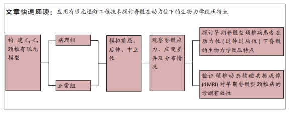
目的:探讨早期脊髓型颈椎病患者在动力位(过伸过屈位)下脊髓的生物力学致压特点,验证颈椎dMRI对早期脊髓型颈椎病诊断的有效性。
方法:回顾性分析2022年1-6月在广西中医药大学第一附属医院骨科就诊并拍摄过颈段dMRI患者的病历资料,共纳入16例,分为2组,病理组为8例以黄韧带肥厚为主要征象的早期脊髓型颈椎病患者,男5例,女3例;正常组为8例正常退变人群,男4例,女4例。所有患者均已拍摄颈椎CT平扫、MRI平扫、dMRI平扫。此次研究分为以下3个部分实验进行阐述:①采集两组受试者dMRI图像的DCOM数据,并采集其颈椎CT、中立位MRI图像DCOM数据了解两组受试者在中立位下的骨质及软组织情况;②基于MRI及CT平扫的图像DCOM数据,使用逆向工程软件分别建模正常退变人群、脊髓型颈椎病早期患者的下颈椎(C3-7)三维有限元模型,分析脊髓、硬膜后部的等效应力、等效弹性应变数值,观察应力、应变分布情况;③得出应力、应变数据后进行组间数据对比,分析出早期脊髓型颈椎病在动力位下脊髓致压的力学特点,验证dMRI对早期脊髓型颈椎病诊断的有效性。

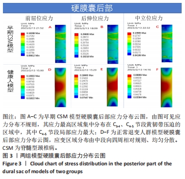
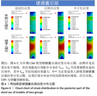
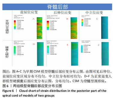
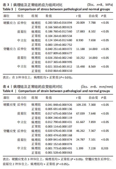
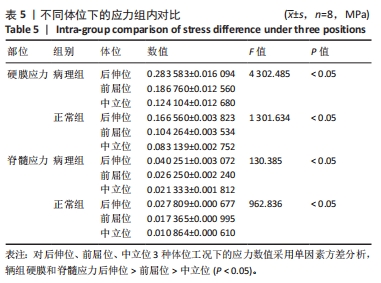
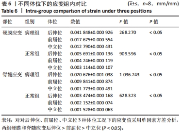
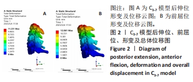
结果与结论:①两组受试者下颈椎(C3-7)模型在模拟后伸、前屈、中立位时,其脊髓后部的应力、应变数值从大到小依次为后伸位>前屈位>中立位(P < 0.05),应变数值从大到小依次为后伸位>前屈位>中立位(P < 0.05);②与正常退变人群模型比较,病理组模型在前屈、后伸2种自由度下,其脊髓的等效应力、应变均较正常组模型增大(P < 0.05),且脊髓后部应力、应变分布区域不规则;③在中立位下,两组模型脊髓的应变数值无明显差异(P > 0.05),应变分布区域均匀规则;④提示在颈椎后伸位状态下,以黄韧带肥厚为主要征象的早期脊髓型颈椎病患者硬膜囊和脊髓后部均受压变形,脊髓的受压形变程度明显大于前屈位和中立位;中立位状态下,早期脊髓型颈椎病患者硬膜受压变形,脊髓无明显形变征象;此次研究从生物力学的角度验证了dMRI对早期脊髓型颈椎病诊断的有效性。

https://orcid.org/0009-0004-6509-3874 (李承蔚)

中国组织工程研究杂志出版内容重点:人工关节;骨植入物;脊柱;骨折;内固定;数字化骨科;组织工程

关键词: 脊髓型颈椎病, 动态MRI(dMRI), 动力位, 早期诊断, 生物力学

Abstract: BACKGROUND: Cervical spondylotic myelopathy is a progressive disease leading to dysfunction in the middle-aged and elderly, and early diagnosis is difficult. In recent years, some clinical scholars have found that dynamic magnetic resonance imaging technology can detect spinal cord compression in a dynamic position earlier, but its specific biomechanical mechanism needs to be clarified. 
OBJECTIVE: To investigate the biomechanical compression characteristics of early cervical spondylotic myelopathy in hyperextension and flexion position, and to verify the effectiveness of dynamic magnetic resonance imaging in the diagnosis of early cervical spondylotic myelopathy. 
METHODS: A retrospective analysis was made on the patients who underwent cervical dynamic magnetic resonance imaging in the Department of Orthopedics of First Affiliated Hospital of Guangxi University of Chinese Medicine from January to June 2022. 16 subjects were selected and divided into two groups. The pathological group included 8 patients with early cervical spondylotic myelopathy with hypertrophy of ligamentum flavum as the main sign, with 5 male patients and 3 female patients. The normal group included 8 normal degenerative people, with 4 male patients and 4 female patients. All patients were photographed with cervical CT plain scan, magnetic resonance imaging plain scan, and dynamic magnetic resonance imaging plain scan. This study was divided into the following three parts: (1) collect the dynamic magnetic resonance imaging image DCOM data of two groups of subjects, and collect the cervical vertebra CT and neutral magnetic resonance imaging image DCOM data to understand the bone and soft tissue of the two groups of subjects in the neutral position. (2) Based on the DCOM data of magnetic resonance imaging and CT plain scan, the three-dimensional finite element models of lower cervical vertebra (C3-7) of normal degenerative population and early cervical spondylotic myelopathy patients were established by reverse engineering software. The equivalent stress and equivalent elastic strain of the spinal cord and posterior dura were analyzed, and the distribution of stress and strain was observed. (3) After obtaining the stress and strain data, the data between groups were compared to analyze the mechanical characteristics of spinal cord compression caused by early cervical spondylotic myelopathy in a dynamic position and to verify the effectiveness of dynamic magnetic resonance imaging in the diagnosis of early cervical spondylotic myelopathy.
RESULTS AND CONCLUSION: (1) When simulating the posterior extension, flexion and neutral position of the lower cervical vertebrae (C3-7) in the two groups, the values of stress and strain in the posterior part of the spinal cord were in the following order: extension > flexion > neutral (P < 0.05). The strain values from large to small were as follows: extension > flexion > neutral (P < 0.05). (2) Compared with the normal degenerative population model, the equivalent stress and strain of the spinal cord in the pathological group were higher than those in the normal group under two degrees of freedom of flexion and extension (P < 0.05). The distribution area of stress and strain in the posterior part of the spinal cord was irregular. (3) In the neutral position, there was no significant difference in the strain value of the spinal cord between the two groups (P > 0.05), and the strain distribution was uniform and regular. (4) It is indicated that in the cervical extension position, the dural sac and the posterior part of the spinal cord were compressed and deformed in the early cervical spondylotic myelopathy patients with the hypertrophy of ligamentum flavum as the main sign, and the degree of compression deformation of the spinal cord was significantly higher than that in the anterior flexion position and neutral position. In the neutral position, there were no obvious signs of spinal cord deformation in patients with early cervical spondylotic myelopathy. This study verified the role of dynamic magnetic resonance imaging in the diagnosis of early cervical spondylotic myelopathy from the point of view of biomechanics.

Key words: cervical spondylotic myelopathy, dynamic magnetic resonance imaging, dynamic position, early diagnosis, biomechanics

中图分类号: