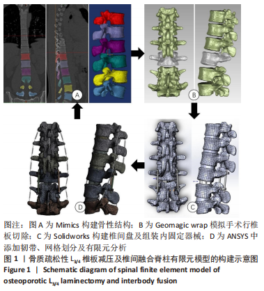
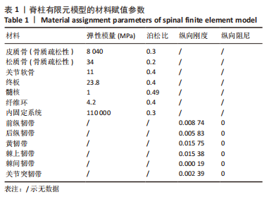
[1] 中华医学会骨质疏松和骨矿盐疾病分会. 原发性骨质疏松症诊疗指南(2017)[J]. 中国全科医学,2017,20(32):3963-3982.
[2] 魏见伟,陈龙伟,姜良海,等. 退变性腰椎滑脱发病的相关因素探讨[J]. 中国矫形外科杂志,2021,29(2):131-134.
[3] TOBERT DG, DAVIS BJ, ANNIS P, et al. The impact of the lordosis distribution index on failure after surgical treatment of adult spinal deformity. Spine J. 2020;20(8):1261-1266.
[4] WANG D, LI Y, YIN H, et al. Three-dimensional finite element analysis of optimal distribution model of vertebroplasty. Ann Palliat Med. 2020;9(3): 1062-1072.
[5] ALY AH, ALY AH, LAI EK, et al. In Vivo Image-Based 4D Modeling of Competent and Regurgitant Mitral Valve Dynamics. Exp Mech. 2021;61(1): 159-169.
[6] LIU Z, WU L, YANG J, et al. Thoracic aorta stent grafts design in terms of biomechanical investigations into flexibility. Math Biosci Eng. 2020;18(1): 800-816.
[7] BELYTSCHKO T, KULAK RF, SCHULTZ AB, et al. Finite element stress analysis of an intervertebral disc. J Biomech. 1974;7(3):277-285.
[8] ZHOU F, YANG S, LIU J, et al. Finite element analysis comparing short-segment instrumentation with conventional pedicle screws and the Schanz pedicle screw in lumbar 1 fractures. Neurosurg Rev. 2020;43(1):301-312.
[9] LO HJ, CHEN HM, KUO YJ, et al. Effect of different designs of interspinous process devices on the instrumented and adjacent levels after double-level lumbar decompression surgery: A finite element analysis. PLoS One. 2020;15(12):e0244571.
[10] MATSUMOTO K, SHAH A, KELKAR A, et al. Biomechanical evaluation of a novel decompression surgery: Transforaminal full-endoscopic lateral recess decompression (TE-LRD). N Am Spine Soc J. 2020;5:100045.
[11] 汪松,马信龙,张弸羽,等. 利用有限元方法测定不同日常动作下坏死股骨头力学变化的研究[J]. 中华骨科杂志,2015,35(9):962-969.
[12] PANJABI MM, WHITE AR. Basic biomechanics of the spine. Neurosurgery. 1980;7(1):76-93.
[13] GOERTZ AR, YANG KH, VIANO DC. Development of a finite element biomechanical whole spine model for analyzing lumbar spine loads under caudocephalad acceleration. Biomed Phys Eng Express. 2020;7(1). doi: 10.1088/2057-1976/abc89a.
[14] YIN JY, GUO LX. Biomechanical analysis of lumbar spine with interbody fusion surgery and U-shaped lumbar interspinous spacers. Comput Methods Biomech Biomed Engin. 2020;26:1-11.
[15] MESSINA C, ACQUASANTA M, RINAUDO L, et al. Short-Term Precision Error of Bone Strain Index, a New DXA-Based Finite Element Analysis Software for Assessing Hip Strength. J Clin Densitom. 2021;24(2):330-337.
[16] RANA M, BISWAS JK, ROY S, et al. Measurement of strain in the rod for lumbar pedicle screw fixation: An experimental and finite element study. Biomed Phys Eng Express. 2020;6(6). doi: 10.1088/2057-1976/abc607.
[17] ROHLMANN A, ZANDER T, BERGMANN G. Spinal loads after osteoporotic vertebral fractures treated by vertebroplasty or kyphoplasty. Eur Spine J. 2006;15(8):1255-1264.
[18] NGUYEN DP, HO BA, THO MC, et al. Reinforcement learning coupled with finite element modeling for facial motion learning. Comput Methods Programs Biomed. 2022;221:106904.
[19] ANA MARÍA MC, JUAN ANTONIO MB. Sacral Prosthesis Substitution as a System of Spinopelvic Reconstruction After Total Sacrectomy: Assessment Using the Finite Element Method. Int J Spine Surg. 2022;16(3):512-520.
[20] HANSRAJ KK, HANSRAJ JA, HANSRAJ MDG, et al. Belly Fat Forces on the Spine: Finite Element Analysis of Belly Fat Forces by Waist Circumference. Surg Technol Int. 2022;40:405-410.
[21] WANG B, KE W, HUA W, et al. Biomechanical evaluation of anterior and posterior lumbar surgical approaches on the adjacent segment: a finite element analysis. Comput Methods Biomech Biomed Engin. 2020;23(14): 1109-1116.
[22] FROST HM. Bone’s mechanostat: a 2003 update. Anat Rec A Discov Mol Cell Evol Biol. 2003;275(2):1081-1101.
[23] 汪松,张弢,马信龙,等. 生物力学因素在股骨头坏死发生发展中的作用研究进展[J]. 山东医药,2015,55(11):89-91.
[24] RAYUDU NM, DIECKMEYER M, LÖFFLER MT, et al. Predicting Vertebral Bone Strength Using Finite Element Analysis for Opportunistic Osteoporosis Screening in Routine Multidetector Computed Tomography Scans-A Feasibility Study. Front Endocrinol (Lausanne). 2021;11:526332.
[25] CAPLLIURE-LLOPIS J, ESCRIVÁ D, BENLLOCH M, et al. Poor Bone Quality in Patients With Amyotrophic Lateral Sclerosis. Front Neurol. 2020;11: 599216.
[26] MARTURELLO DM, VON PFEIL DJF, DÉJARDIN LM. Evaluation of a Feline Bone Surrogate and In Vitro Mechanical Comparison of Small Interlocking Nail Systems in Mediolateral Bending. Vet Comp Orthop Traumatol. 2021; 34(4):223-233.
[27] TADANO S, TANABE H, ARAI S, et al. Lumbar mechanical traction: a biomechanical assessment of change at the lumbar spine. BMC Musculoskelet Disord. 2019;20(1):155.
[28] WARREN JM, MAZZOLENI AP, HEY LA. Development and Validation of a Computationally Efficient Finite Element Model of the Human Lumbar Spine: Application to Disc Degeneration. Int J Spine Surg. 2020;14(4):502-510.
[29] LO HJ, CHEN HM, KUO YJ, et al. Effect of different designs of interspinous process devices on the instrumented and adjacent levels after double-level lumbar decompression surgery: A finite element analysis. PLoS One. 2020;15(12):e0244571.
[30] MATSUKAWA K, YATO Y, IMABAYASHI H. Impact of Screw Diameter and Length on Pedicle Screw Fixation Strength in Osteoporotic Vertebrae: A Finite Element Analysis. Asian Spine J. 2021;15(5):566-574. |