中国组织工程研究 ›› 2024, Vol. 28 ›› Issue (5): 697-705.doi: 10.12307/2023.885
• 水凝胶材料Hydrogel materials • 上一篇 下一篇
负载外泌体水凝胶修饰3D打印支架构建血管化的气道替代物
沈子青1,夏 天1,单一波2,朱睿君1,万昊鑫1,丁 浩1,潘 枢1,赵 军1
- 1苏州大学附属第一医院胸外科,江苏省苏州市 215006;2扬州大学医学院转化学研究院,江苏省扬州市 225000
-
收稿日期:2022-11-24接受日期:2023-01-05出版日期:2024-02-18发布日期:2023-08-16 -
通讯作者:赵军,博士,教授,主任医师,苏州大学附属第一医院胸外科,江苏省苏州市 215006 -
作者简介:沈子青,男,1996年生,江苏省苏州市人,汉族,苏州大学第一临床医学院在读硕士,主要从事组织工程气管替代治疗的研究。 -
基金资助:江苏省自然科学基金青年项目(BK20200196);项目负责人:潘枢;苏州大学企业委托项目(H221026),项目负责人:赵军
Vascularized tracheal substitutes constructed by exosome-load hydrogel-modified 3D printed scaffolds
Shen Ziqing1, Xia Tian1, Shan Yibo2, Zhu Ruijun1, Wan Haoxin1, Ding Hao1, Pan Shu1, Zhao Jun1
- 1Department of Thoracic Surgery, The First Affiliated Hospital of Soochow University, Suzhou 215006, Jiangsu Province, China; 2Institute of Translational Research, College of Medicine, Yangzhou University, Yangzhou 225000, Jiangsu Province, China
-
Received:2022-11-24Accepted:2023-01-05Online:2024-02-18Published:2023-08-16 -
Contact:Zhao Jun, PhD, Professor, Chief physician, Department of Thoracic Surgery, The First Affiliated Hospital of Soochow University, Suzhou 215006, Jiangsu Province, China -
About author:Shen Ziqing, Master candidate, Department of Thoracic Surgery, The First Affiliated Hospital of Soochow University, Suzhou 215006, Jiangsu Province, China -
Supported by:Natural Science Foundation for the Youth of Jiangsu Province, No. BK20200196 (to PS); Enterprise Commissioned Project of Soochow University, No. H221026 (to ZJ)
摘要:

文题释义:
外泌体:是一种由细胞分泌的包含有核酸、脂质和蛋白质的纳米囊泡状结构,在细胞间的交流中发挥着不可或缺的调节作用。甲基丙烯酰化透明质酸水凝胶:透明质酸是由D-葡萄糖醛酸和N-乙酰-D-氨基葡萄糖作为双糖结构单元的天然糖胺多糖聚合物,在细胞增殖、分化、形态发生和伤口愈合等生物学过程中发挥重要作用。在其分子链上接枝丙烯酸基团获得甲基丙烯酰化透明质酸,使其进一步具有光固化特性。
背景:对于长段气管缺损的替代治疗,尽管近年来组织工程研究取得了一定的进展,但仍然不尽完善,其中最大的难点就是不能快速实现气管替代物的血运重建。
目的:初步探索经负载外泌体水凝胶修饰的聚己内酯支架构建可快速血管化气管替代物的潜力。方法:提取SD大鼠骨髓间充质干细胞来源外泌体,制备甲基丙烯酰化透明质酸溶液,将外泌体溶液与甲基丙烯酰化透明质酸溶液按照体积比1∶1混合,紫外光照射5 min制备负载外泌体的甲基丙烯酰化透明质酸水凝胶。检测未负载外泌体水凝胶的降解情况,负载外泌体水凝胶外泌体的控制释放情况。采用3D打印法制备聚己内酯支架,将单纯的甲基丙烯酰化透明质酸溶液与负载外泌体的甲基丙烯酰化透明质酸水溶液分别滴加至支架表面,紫外光照射后获得水凝胶修饰的支架与外泌体修饰的支架。将30只SD大鼠采用随机数字表法分为3组,每组10只,分别于皮下包埋单纯的支架、水凝胶修饰的支架与外泌体修饰的支架,术后30 d取出各组支架及周围组织,通过苏木精-伊红及Masson染色观察新生血管生成情况,免疫荧光检测CD31的表达。
结果与结论:①随着时间的推移,水凝胶逐渐降解,并且包封于水凝胶中的外泌体逐渐释放,可持续释放至30 d以上,并且外泌体的释放速率快于水凝胶自身的降解速率,浸泡30 d时仍有近20%的外泌体未被释放;②扫描电镜下可见,单纯的聚己内酯支架表面粗糙,经水凝胶修饰后,支架孔隙之间覆盖一层凝胶物质,支架表面变得光滑、致密;③大鼠皮下包埋30 d后,苏木精-伊红及Masson染色显示,与水凝胶修饰支架组相比,外泌体修饰支架组支架内部可见更多的新生血管形成,两组支架表面的水凝胶未完全降解;免疫荧光染色显示,外泌体修饰支架组CD31表达高于水凝胶修饰支架组(P < 0.000 1);④结果表明,甲基丙烯酰化透明质酸水凝胶可以作为外泌体的控释载体,经负载外泌体甲基丙烯酰化透明质酸水凝胶修饰后的3D打印聚己内酯支架,具有良好的生物相容性,且具有促进新生血管形成的潜力。
https://orcid.org/0000-0003-3916-6983(沈子青)
中国组织工程研究杂志出版内容重点:生物材料;骨生物材料;口腔生物材料;纳米材料;缓释材料;材料相容性;组织工程
中图分类号:
引用本文
沈子青, 夏 天, 单一波, 朱睿君, 万昊鑫, 丁 浩, 潘 枢, 赵 军. 负载外泌体水凝胶修饰3D打印支架构建血管化的气道替代物[J]. 中国组织工程研究, 2024, 28(5): 697-705.
Shen Ziqing, Xia Tian, Shan Yibo, Zhu Ruijun, Wan Haoxin, Ding Hao, Pan Shu, Zhao Jun. Vascularized tracheal substitutes constructed by exosome-load hydrogel-modified 3D printed scaffolds[J]. Chinese Journal of Tissue Engineering Research, 2024, 28(5): 697-705.
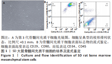
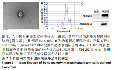
2.2 外泌体的鉴定结果 采用BCA法检测外泌体中的蛋白质量浓度为0.503 65 μg/μL。透射电镜下可见,骨髓间充质干细胞来源的外泌体具有明显的脂质双层膜圆形结构,直径约为150 nm,见图2A,即为外泌体。结果表示该法提取的外泌体符合广泛接受的标准特征。粒径检测结果表明,所提取及纯化外泌体悬液中的颗粒平均直径为188.1 nm,所提取外泌体的浓度约为1.0×10¹² L-1,见图2B。Western blot检测结果显示,骨髓间充质干细胞来源的外泌体高表达标志蛋白TSG101及Alix,骨髓间充质干细胞并不表达或仅微弱表达以上蛋白,见图2C。
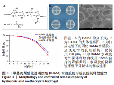
2.3 HAMA水凝胶的形貌及控释能力检测结果 图3A为HAMA的化学式,其大体形态如图3B所示。扫描电镜下可见固化HAMA水凝胶呈现光滑的孔状结构,见图3C所示。图3D显示了HAMA水凝胶对外泌体的控释能力及HAMA本身的降解特性。可以看出,随着时间的延长,外泌体被逐渐释放,当浸泡到30 d时,经分析计算,仍有约20%的外泌体保存于固化HAMA水凝胶中。同样,随着时间的延长,HAMA水凝胶自身也在降解,至30 d时剩约30%的HAMA,说明水凝胶的降解速率慢于外泌体的释放速率。该结果表明,通过固化HAMA水凝胶来实现对外泌体的控制释放的方法是可行的。
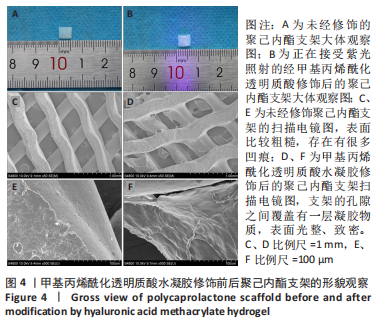
2.4 3D打印多孔聚己内酯支架的形貌观察结果 实验打印出直径约200 μm、大小约0.5 cm×0.5 cm的聚己内酯支架,见图4A;在纯聚己内酯支架上滴加少量HAMA水凝胶溶液,经紫灯照射5 min后等待其固化,见图4B。扫描电镜下可以观察到,原先的聚己内酯材料表面比较粗糙,存在有很多凹痕,见图4C、E。经HAMA修饰处理后,支架的孔隙之间覆盖有一层凝胶物质,见图4D;高倍镜下可见材料之前变得光整、致密,原先的凹痕基本消失,见图4F。
2.5 气管替代物大鼠皮下包埋实验结果
2.5.1 实验动物数量分析 30只大鼠全部进入结果分析。
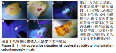
2.5.2 大鼠术中情况、术后生存情况及标本收获 构建大鼠皮囊模型后,将补片植入皮下,术中出血少,采取间断缝合法缝合伤口。单纯支架组切开皮肤后立即将单纯聚己内酯支架植入皮下。将提前制备好的HAMA及负载外泌体的HAM水凝胶溶液抽吸于2 mL避光针筒中,紫光短暂照射针尖,使其达到滴出便可固化状态,将其滴加于聚己内酯支架表面,分别获得水凝胶修饰的支架与外泌体修饰的支架,植入皮下,见图5。
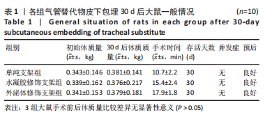
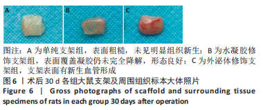
各组大鼠术后均未出现免疫排斥反应或其他并发症,术后一般情况均良好,如表1所示。于术后30 d时收获大体标本,见图6,单纯支架组表面粗糙,未见明显组织新生;水凝胶修饰支架组可见表面覆盖凝胶仍未完全降解,形态良好;外泌体修饰支架组支架表面有明显的新生血管形成。
2.5.3 各组标本组织学分析结果 由于聚己内酯材料是酯溶性的,因此在制蜡过程中会发生溶解,因而后续实验仅进行了水凝胶修饰支架及外泌体修饰支架组的切片组织学分析。
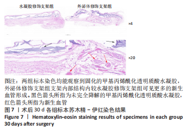
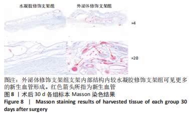
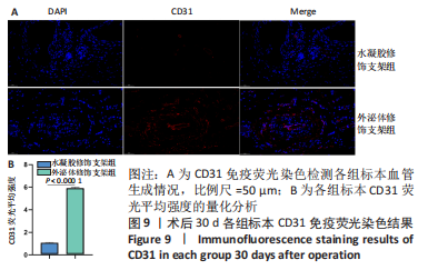
术后30 d的苏木精-伊红及Masson染色可以观察到,外泌体修饰支架组支架内部结构内较水凝胶修饰支架组可见更多的新生血管形成,两组标本染色均能观察到固化的HAMA水凝胶,支架表面的HAMA于30 d时仍未完全降解,见图7,8,不仅提高了气管替代物支架的生物相容性,还实现了对外泌体的控制释放,使其更精准和更长时间的作用于靶点。免疫荧光染色显示,外泌体修饰支架组CD31表达高于水凝胶修饰支架组(P < 0.000 1),见图9。
2.6 支架的生物相容性 聚己内酯是一种非免疫原性、非致癌性、可生物降解、降解产物无毒、生物相容性良好的疏水材料[47]。透明质酸是动物组织的细胞外基质的成分,具有良好的锁水保湿性能,在细胞增殖、分化、形态发生、炎症和伤口愈合中发挥重要作用[48]。外泌体是细胞分泌的纳米级膜囊泡物质,拥有良好的稳定性和极低的免疫原性[49]。该研究将经负载外泌体HAMA水凝胶修饰的聚己内酯支架种植到大鼠皮下,均未出现皮肤溃疡、腹泻、体质量下降等表现,未见明显的移植物抗宿主反应,具有良好的生物相容性。

| [1] LEE H, MARIN-ARAUJO AE, AOKI FG, et al. Computational fluid dynamics for enhanced tracheal bioreactor design and long-segment graft recellularization. Sci Rep. 2021;11(1):1187. [2] CHOI JS, LEE MS, KIM J, et al. Hyaluronic Acid Coating on Hydrophobic Tracheal Scaffold Enhances Mesenchymal Stem Cell Adhesion and Tracheal Regeneration. Tissue Eng Regen Med. 2021;18:225-233. [3] XU C, MA Y, HUANG H, et al. A Review of Woven Tracheal Stents: Materials, Structures, and Application. J Funct Biomater. 2022;13(3):96. [4] YU YS, AHN CB, SON KH, et al. Motility Improvement of Biomimetic Trachea Scaffold via Hybrid 3D-Bioprinting Technology. Polymers (Basel) 2021;13(6):971. [5] SORIANO L, KHALID T, WHELAN D, et al. Development and clinical translation of tubular constructs for tracheal tissue engineering: a review. Eur Respir Rev. 2021;30:(162):210154. [6] VRANCKX JJ, DELAERE P. The current status and outlook of trachea transplantation. Curr Opin Organ Transplant. 2020;25:601-608. [7] GAO B, JING H, GAO M, et al. Long-segmental tracheal reconstruction in rabbits with pedicled Tissue-engineered trachea based on a 3D-printed scaffold. Acta Biomater. 2019;97:177-186. [8] Kim IG, Park SA, Lee SH, et al. Transplantation of a 3D-printed tracheal graft combined with iPS cell-derived MSCs and chondrocytes. Sci Rep. 2020;10(1):4326. [9] XIA D, JIN D, WANG Q, et al. Tissue-engineered trachea from a 3D-printed scaffold enhances whole-segment tracheal repair in a goat model. J Tissue Eng Regen Med. 2019;13:694-703. [10] YAO ZY, FENG BW, LIU CS, et al. The Application of a Bone Marrow Mesenchymal Stem Cell Membrane in the Vascularization of a Decellularized Tracheal Scaffold. Stem Cells Int. 2021;2021:6624265. [11] XIANG Y, WANG W, GAO Y, et al. Production and Characterization of an Integrated Multi-Layer 3D Printed PLGA/GelMA Scaffold Aimed for Bile Duct Restoration and Detection. Front Bioeng Biotechnol. 2020;8:971. [12] GRIGORYAN B, PAULSEN SJ, CORBETT DC, et al. Multivascular networks and functional intravascular topologies within biocompatible hydrogels. Science. 2019;364:458-464. [13] UDELSMAN B, MATHISEN DJ, OTT HC. A reassessment of tracheal substitutes-a systematic review. Ann Cardiothorac Surg. 2018;7:175-182. [14] HUO Y, XU Y, WU X, et al. Functional Trachea Reconstruction Using 3D-Bioprinted Native-Like Tissue Architecture Based on Designable Tissue-Specific Bioinks. Adv Sci (Weinh). 2022;9:e2202181. [15] PARK JY, RYU H, LEE B, et al. Development of a functional airway-on-a-chip by 3D cell printing. Biofabrication. 2018;11:015002. [16] DHASMANA A, SINGH A, RAWAL S. Biomedical grafts for tracheal tissue repairing and regeneration “Tracheal tissue engineering: an overview”. J Tissue Eng Regen Med. 2020; 14:653-672. [17] MAUGHAN EF, BUTLER CR, CROWLEY C, et al. A comparison of tracheal scaffold strategies for pediatric transplantation in a rabbit model. Laryngoscope. 2017;127:E449-e457. [18] REHMANI SS, AL-AYOUBI AM, AYUB A, et al. Three-Dimensional-Printed Bioengineered Tracheal Grafts: Preclinical Results and Potential for Human Use. Ann Thorac Surg. 2017; 104:998-1004. [19] LEE JS, PARK J, SHIN DA, et al. Characterization of the biomechanical properties of canine trachea using a customized 3D-printed apparatus. Auris Nasus Larynx. 2019;46:407-416. [20] BAE SW, LEE KW, PARK JH, et al. 3D Bioprinted Artificial Trachea with Epithelial Cells and Chondrogenic-Differentiated Bone Marrow-Derived Mesenchymal Stem Cells. Int J Mol Sci. 2018;19(6):1624. [21] MACHINO R, MATSUMOTO K, TANIGUCHI D, et al. Replacement of Rat Tracheas by Layered, Trachea-Like, Scaffold-Free Structures of Human Cells Using a Bio-3D Printing System. Adv Healthc Mater. 2019;8:e1800983. [22] KATAGIRI W, TAKEUCHI R, SAITO N, et al. Migration and phenotype switching of macrophages at early-phase of bone-formation by secretomes from bone marrow derived mesenchymal stem cells using rat calvaria bone defect model. J Dent Sci. 2022;17:421-429. [23] AL-QADHI G, ABOUSHADY I, AL-SHARABI N. The Gingiva from the Tissue Surrounding the Bone to the Tissue Regenerating the Bone: A Systematic Review of the Osteogenic Capacity of Gingival Mesenchymal Stem Cells in Preclinical Studies. Stem Cells Int. 2021; 2021:6698100. [24] NASSERI MALEKI S, ABOUTALEB N, NAZARINIA D, et al. Conditioned medium obtained from human amniotic membrane-derived mesenchymal stem cell attenuates heart failure injury in rats. Iran J Basic Med Sci. 2019;22:1253-1258. [25] SISSUNG TM, FIGG WD. Stem cell clinics: risk of proliferation. Lancet Oncol. 2020;21:205-206. [26] BIAN X, MA K, ZHANG C, et al. Therapeutic angiogenesis using stem cell-derived extracellular vesicles: an emerging approach for treatment of ischemic diseases. Stem Cell Res Ther. 2019;10:158. [27] JESKE R, BEJOY J, MARZANO M, et al. Human Pluripotent Stem Cell-Derived Extracellular Vesicles: Characteristics and Applications. Tissue Eng Part B Rev. 2020;26:129-144. [28] CAO J, ZHANG M, XIE F, et al. Exosomes in head and neck cancer: Roles, mechanisms and applications. Cancer Lett. 2020;494:7-16. [29] ROCCARO AM, SACCO A, MAISO P, et al. BM mesenchymal stromal cell-derived exosomes facilitate multiple myeloma progression. J Clin Invest. 2013;123:1542-1555. [30] WU D, CHANG X, TIAN J, et al. Bone mesenchymal stem cells stimulation by magnetic nanoparticles and a static magnetic field: release of exosomal miR-1260a improves osteogenesis and angiogenesis. J Nanobiotechnology. 2021;19:209. [31] HUANG Y, HE B, WANG L, et al. Bone marrow mesenchymal stem cell-derived exosomes promote rotator cuff tendon-bone healing by promoting angiogenesis and regulating M1 macrophages in rats. Stem Cell Res Ther. 2020;11:496. [32] LIAO W, NING Y, XU HJ, et al. BMSC-derived exosomes carrying microRNA-122-5p promote proliferation of osteoblasts in osteonecrosis of the femoral head. Clin Sci (Lond). 2019;133:1955-1975. [33] LIANG B, LIANG JM, DING JN, et al. Dimethyloxaloylglycine-stimulated human bone marrow mesenchymal stem cell-derived exosomes enhance bone regeneration through angiogenesis by targeting the AKT/mTOR pathway. Stem Cell Res Ther. 2019;10:335. [34] XU H, WANG Z, LIU L, et al. Exosomes derived from adipose tissue, bone marrow, and umbilical cord blood for cardioprotection after myocardial infarction. J Cell Biochem. 2020;121:2089-2102. [35] SHABBIR A, COX A, RODRIGUEZ-MENOCAL L, et al. Mesenchymal Stem Cell Exosomes Induce Proliferation and Migration of Normal and Chronic Wound Fibroblasts, and Enhance Angiogenesis In Vitro. Stem Cells Dev. 2015;24:1635-1647. [36] ZENG T, YUAN P, LIANG L, et al. Cartilaginous Extracellular Matrix Enriched with Human Gingival Mesenchymal Stem Cells Derived “Matrix Bound Extracellular Vesicles” Enabled Functional Reconstruction of Tracheal Defect. Adv Sci (Weinh). 2022;9:e2102735. [37] HUA T, YANG M, SONG H, et al. Huc-MSCs-derived exosomes attenuate inflammatory pain by regulating microglia pyroptosis and autophagy via the miR-146a-5p/TRAF6 axis. J Nanobiotechnology. 2022;20:324. [38] GUO L, CHEN Y, FENG X, et al. Oxidative stress-induced endothelial cells-derived exosomes accelerate skin flap survival through Lnc NEAT1-mediated promotion of endothelial progenitor cell function. Stem Cell Res Ther. 2022;13:325. [39] WANG B, WANG ZM, JI JL, et al. Macrophage-Derived Exosomal Mir-155 Regulating Cardiomyocyte Pyroptosis and Hypertrophy in Uremic Cardiomyopathy. JACC Basic Transl Sci. 2020;5:148-166. [40] YERNENI SS, LATHWAL S, CUTHBERT J, et al. Controlled Release of Exosomes Using Atom Transfer Radical Polymerization-Based Hydrogels. Biomacromolecules. 2022;23:1713-1722. [41] YANG J, CHEN Z, PAN D, et al. Umbilical Cord-Derived Mesenchymal Stem Cell-Derived Exosomes Combined Pluronic F127 Hydrogel Promote Chronic Diabetic Wound Healing and Complete Skin Regeneration. Int J Nanomedicine. 2020;15:5911-5926. [42] KAWANO Y, PATRULEA V, SUBLET E, et al. Wound Healing Promotion by Hyaluronic Acid: Effect of Molecular Weight on Gene Expression and In Vivo Wound Closure. Pharmaceuticals (Basel) 2021;14(4):301. [43] XIA H, ZHAO D, ZHU H, et al. Lyophilized Scaffolds Fabricated from 3D-Printed Photocurable Natural Hydrogel for Cartilage Regeneration. ACS Appl Mater Interfaces. 2018;10: 31704-31715. [44] CHEN P, ZHENG L, WANG Y, et al. Desktop-stereolithography 3D printing of a radially oriented extracellular matrix/mesenchymal stem cell exosome bioink for osteochondral defect regeneration. Theranostics. 2019;9:2439-2459. [45] WANG C, DU H, HOU J, et al. Chaihulonggumulitang Shows Psycho-cardiology Therapeutic Effects on Acute Myocardial Infarction by Enhancing Bone Marrow Mesenchymal Stem Cells Mobilization. Sci Rep. 2018;8:3724. [46] PAN S, ZHONG Y, SHAN Y, et al. Selection of the optimum 3D-printed pore and the surface modification techniques for tissue engineering tracheal scaffold in vivo reconstruction. J Biomed Mater Res A. 2019;107:360-370. [47] HUANG HY, CHEN LQ, SUN W, et al. Collagenase IV and clusterin-modified polycaprolactone-polyethylene glycol nanoparticles for penetrating dense tumor tissues. Theranostics. 2021;11:906-924. [48] INUBUSHI T, NAKANISHI Y, ABE M, et al. The cell surface hyaluronidase TMEM2 plays an essential role in mouse neural crest cell development and survival. PLoS Genet. 2022; 18(7):e1009765. [49] WAN Z, GAN X, MEI R, et al. ROS triggered local delivery of stealth exosomes to tumors for enhanced chemo/photodynamic therapy. J Nanobiotechnology 2022;20(1):385. [50] BATIOGLU-KARAALTIN A, OVALI E, KARAALTIN MV, et al. Decellularization of Trachea With Combined Techniques for Tissue-Engineered Trachea Transplantation. Clin Exp Otorhinolaryngol. 2019;12(1):86-94. [51] KIM H, LEE JY, HAN H, et al. Improved chondrogenic performance with protective tracheal design of Chitosan membrane surrounding 3D-printed trachea. Sci Rep. 2021;11(1):9258. [52] LIU L, DHARMADHIKARI S, SPECTOR BM, et al. Tissue-engineered composite tracheal grafts create mechanically stable and biocompatible airway replacements. J Tissue Eng. 2022;13:20417314221108791. [53] MASSOUMI B, HATAMZADEH M, FIROUZI N, et al. Electrically conductive nanofibrous scaffold composed of poly(ethylene glycol)-modified polypyrrole and poly(ε-caprolactone) for tissue engineering applications. Mater Sci Eng C Mater Biol Appl. 2019;98:300-310. [54] LUO Y, GUI R. Circulating exosomal circFoxp1 confers cisplatin resistance in epithelial ovarian cancer cells. J Gynecol Oncol 2020;31(5):e75. [55] ZHOU Z, WANG R, WANG J, et al. Melatonin pretreatment on exosomes: Heterogeneity, therapeutic effects, and usage. Front Immunol 2022;13:933736. [56] WEI Q, WANG Y, MA K, et al. Extracellular Vesicles from Human Umbilical Cord Mesenchymal Stem Cells Facilitate Diabetic Wound Healing Through MiR-17-5p-mediated Enhancement of Angiogenesis. Stem Cell Rev Rep. 2022;18:1025-1040. [57] XIAO S, XIAO C, MIAO Y, et al. Human acellular amniotic membrane incorporating exosomes from adipose-derived mesenchymal stem cells promotes diabetic wound healing. Stem Cell Res Ther. 2021;12(1):255. [58] ZHANG X, JIANG Y, HUANG Q, et al. Exosomes derived from adipose-derived stem cells overexpressing glyoxalase-1 protect endothelial cells and enhance angiogenesis in type 2 diabetic mice with limb ischemia. Stem Cell Res Ther. 2021;12(1):403. [59] MAO Q, LIANG XL, ZHANG CL, et al. LncRNA KLF3-AS1 in human mesenchymal stem cell-derived exosomes ameliorates pyroptosis of cardiomyocytes and myocardial infarction through miR-138-5p/Sirt1 axis. Stem Cell Res Ther. 2019;10(1):393. [60] NI J, LIU X, YIN Y, et al. Exosomes Derived from TIMP2-Modified Human Umbilical Cord Mesenchymal Stem Cells Enhance the Repair Effect in Rat Model with Myocardial Infarction Possibly by the Akt/Sfrp2 Pathway. Oxid Med Cell Longev. 2019;2019:1958941. [61] ZHU D, WANG Y, THOMAS M, et al. Exosomes from adipose-derived stem cells alleviate myocardial infarction via microRNA-31/FIH1/HIF-1α pathway. J Mol Cell Cardiol. 2022; 162:10-19. [62] ABBASS MMS, EL-RASHIDY AA, SADEK KM, et al. Hydrogels and Dentin-Pulp Complex Regeneration: From the Benchtop to Clinical Translation. Polymers (Basel). 2020;12(12): 2935. [63] PAN S, SHEN Z, XIA T, et al. Hydrogel modification of 3D printing hybrid tracheal scaffold to construct an orthotopic transplantation. Am J Transl Res. 2022;14:2910-2925. [64] YAZDANI M, SHAHDADFAR A, JACKSON CJ, et al. Hyaluronan-Based Hydrogel Scaffolds for Limbal Stem Cell Transplantation: A Review. Cells. 2019;8(3):245. [65] WIKLANDER OPB, BRENNAN M, LÖTVALL J, et al. Advances in therapeutic applications of extracellular vesicles. Sci Transl Med. 2019;11(492):eaav8521. |
| [1] | 王梦晗, 齐 涵, 张 元, 陈言智. 3种3D打印模型辅助治疗Robinson ⅡB2型锁骨骨折[J]. 中国组织工程研究, 2024, 28(9): 1403-1408. |
| [2] | 杨玉芳, 杨芷姗, 段棉棉, 刘毅恒, 唐正龙, 王 宇. 促红细胞生成素在骨组织工程中的应用及前景[J]. 中国组织工程研究, 2024, 28(9): 1443-1449. |
| [3] | 陈凯佳, 刘景云, 曹 宁, 孙建波, 周 燕, 梅建国, 任 强. 组织工程技术在股骨头坏死治疗中的应用及前景[J]. 中国组织工程研究, 2024, 28(9): 1450-1456. |
| [4] | 梅静怡, 刘 江, 肖 聪, 刘 鹏, 周浩浩, 林展翼. 组织工程血管构建过程中平滑肌细胞增殖变化及代谢模式[J]. 中国组织工程研究, 2024, 28(7): 1043-1049. |
| [5] | 王姗姗, 舒 晴, 田 峻. 物理因子促进干细胞的成骨分化[J]. 中国组织工程研究, 2024, 28(7): 1083-1090. |
| [6] | 刘瀚峰, 王晶晶, 余云生. 人造外泌体治疗心肌梗死:应用现状及前景[J]. 中国组织工程研究, 2024, 28(7): 1118-1123. |
| [7] | 马树微, 何 生, 韩 冰, 张缭云. 间充质干细胞来源外泌体治疗动物急性肝衰竭的Meta分析[J]. 中国组织工程研究, 2024, 28(7): 1137-1142. |
| [8] | 宁天亮, 王 坤, 王领彪, 韩鹏飞. 基于三点力原理对足内翻矫形器矫正效果的有限元分析[J]. 中国组织工程研究, 2024, 28(6): 891-899. |
| [9] | 张之栋, 祁家龙, 裴少保, 马 力, 王善松, 刘艺明. 3D打印导板技术联合多次去旋转治疗重度僵硬性脊柱侧凸[J]. 中国组织工程研究, 2024, 28(6): 922-926. |
| [10] | 王 武, 樊晓磊, 谢 杰, 胡懿郃, 曾 敏. 羟基磷灰石-聚乙烯醇/胶原-壳聚糖-明胶复合水凝胶修复兔骨软骨缺损[J]. 中国组织工程研究, 2024, 28(5): 682-689. |
| [11] | 张 娅, 牟秋菊, 王自林, 刘宏杰, 祝丽丽. 负载富血小板血浆的水凝胶促进糖尿病大鼠创面愈合[J]. 中国组织工程研究, 2024, 28(5): 690-696. |
| [12] | 朱礼威, 王江玥, 白 丁. 纳米复合甲基丙烯酰明胶水凝胶在不同骨缺损环境中应用的价值[J]. 中国组织工程研究, 2024, 28(5): 753-758. |
| [13] | 陈小芳, 郑国爽, 李茂源, 于炜婷. 可注射海藻酸钠水凝胶的制备及应用[J]. 中国组织工程研究, 2024, 28(5): 789-794. |
| [14] | 王嘉旎, 陈俊宇. 金属离子促血管生成机制及在骨组织工程中的应用[J]. 中国组织工程研究, 2024, 28(5): 804-812. |
| [15] | 杨雨晴, 陈志宇. 早期短暂M1巨噬细胞在骨组织工程中的作用及应用[J]. 中国组织工程研究, 2024, 28(4): 594-601. |
3D打印技术有着生产周期短、高效、匹配度高、所生产支架力学性能好且能满足个体化需求的优点,是一种快速成型及逐层打印技术,该技术可以把多种材料混合并进行单次或多次打印以构建所需要的气管结构[15]。常用的支架材料主要分为天然材料和合成材料,天然材料包括壳聚糖、海藻酸盐、脱细胞细胞外基质等,而常用的合成材料包括聚已内酯、聚氨酯、聚乙二醇、聚乙醇酸等[16-17]。前沿研究发现,天然材料制备的气管替代物往往具有生物相容性佳、免疫原性低、可为移植处细胞增殖分化提供良好微环境等优点,但存在生物力学性能不足的缺点,移植术后短期内便可出现气管管腔塌陷,进而引起气道堵塞与窒息而导致移植失败[16,18]。为进一步制备出生物力学性能良好的组织工程气管替代物,LEE等[19]使用聚己内酯作为打印材料结合3D打印技术制备出了一种圆柱形气管支架,后续动物实验证明其力学性能与原生气管类似,可以满足气管替代的长期机械支撑,但由于其不能快速重建新生血管及上皮形成不足和黏液阻塞而导致失败。虽然聚己内酯支架有着可降解吸收、可打印性、无毒性、优异的机械性能、良好的孔隙率等优点,但存在细胞亲和力不足的劣势,因此需要对聚己内酯支架表面进行修饰来进一步提高其生物相容性。
近年来,得益于干细胞学的发展,骨髓间充质干细胞移植为治疗长段气管缺损提供了新的思路与方向[20-21]。骨髓间充质干细胞作为组织工程中常用的明星“种子细胞”,具有易获取、增殖旺盛、可定向诱导分化成为多种类型细胞及促进其他细胞增殖和分化等优点[10,22-23]。但单纯干细胞移植疗法也面临诸多难题,如移植部位细胞滞留率和生存率低、耗时长、有免疫排斥反应风险、致瘤性、微环境影响等[24-25],因而限制了其临床应用。而近年研究表明,干细胞可以通过其分泌的细胞因子发挥进一步作用[26]。外泌体作为干细胞旁分泌的重要物质,是包含有核酸、脂质和蛋白质的纳米囊泡状结构[26],并通过转移这些物质至靶细胞而发挥功能,在细胞间的交流中发挥着不可或缺的作用[27-28]。外泌体表现出类似干细胞的促再生特性,且外泌体不含主要组织相容性复合物Ⅰ或主要组织相容性复合物Ⅱ蛋白,因此直接用外泌体治疗可以避免干细胞移植治疗的许多不良反应[29]。近年来,在前沿组织工程研究领域,无论是在骨肩袖撕裂、股骨头坏死、糖尿病伤口愈合及心肌梗死等缺血性疾病,骨髓间充质干细胞衍生的外泌体都显示出其促进血管生成、减轻炎症反应、减少细胞凋亡及促进伤口愈合等能力[30-35]。而在气管组织工程领域,一些研究者已经开始通过应用干细胞衍生的外泌体来构建可快速上皮化的气管替代物,并取得了一定的进展[36]。然而,对于实现气管替代物的血供重建,这种通过使用干细胞外泌体的疗法并没有广泛开展,值得进一步研究。
既往对外泌体的研究往往通过体内静脉注射或局部注射外泌体来进行相关研究[37-39],该方法取得了一定的成果,但存在着外泌体需反复注射、治疗靶点滞留率低、时效短、需求量大等缺点。而气管缺损的修复重建是一个长周期的过程,因此保证替代物中所负载的因子类物质能尽可能地发挥作用十分重要。前沿研究表明,相比于直接注射,凝胶包裹外泌体是一种更有前景的治疗方法。YERNENI等[40]使用自由基聚合水凝胶来控制外泌体的释放,结果表明将外泌体负载在水凝胶网络中可以使外泌体在1个月内缓慢释放。无独有偶,YANG等[41]使用PF-127水凝胶作为间充质干细胞来源外泌体的控释载体,相比单一使用外泌体,这种水凝胶包裹外泌体的方法可进一步促进慢性糖尿病伤口愈合和皮肤完全再生。不同类型的单一水凝胶材料往往有着各自的优势与缺点,如透明质酸是一种天然糖胺多糖聚合物,具有良好的锁水性能,在细胞增殖、分化、形态发生、抗炎症和伤口愈合等许多生物学过程中发挥着重要作用,但其降解速度过快、结构不稳定[42]。为进一步制备出理想的水凝胶载体,XIA等[43]在明胶和透明质酸上接枝甲基丙烯酸,通过整合3D打印和冻干技术获得了具备很好的剪切变稀特性的甲基丙烯酰化明胶和甲基丙烯酰化透明质酸(hyaluronic acid methacrylate,HAMA)复合水凝胶。CHEN等[44]将软骨脱细胞外基质与甲基丙烯酰化明胶混合,并负载从骨髓间充质干细胞提取的外泌体,通过立体光固化成型打印获得复合支架。这种通过对水凝胶进行化学改性而进一步获得的复合水凝胶,在保持原有水凝胶优势的同时延长了其降解周期,增加了稳定性,但缺点是仍然力学性能不佳。因此,将这种负载了外泌体的复合水凝胶进一步修饰在聚己内酯支架上,不仅可以实现对外泌体的良好负载,获到更佳更长的外泌体控释效果,而且获得了具有优异机械性能、良好的保水性、抗感染能力和出色的细胞生物相容性的复合气管支架,具有良好的应用前景。
在此基础上,此次实验将HAMA水凝胶作为骨髓间充质干细胞来源外泌体的载体,对3D打印聚己内酯支架进行进一步修饰,探究其能否促进新生血管形成,为后续长段气管缺损替代治疗提供新的方案与选择。 中国组织工程研究杂志出版内容重点:生物材料;骨生物材料;口腔生物材料;纳米材料;缓释材料;材料相容性;组织工程
1.2 时间及地点 实验于2020年9月至2022年9月在苏州大学附属第一医院基础实验室完成。
1.3 材料
1.3.1 实验动物 40只SPF级健康SD大鼠,雌雄不拘,10周龄左右,体质量300-400 g,均购自昭衍(苏州)新药研究中心有限公司,许可证号:SCXK(苏)2018-0006。大鼠饲养于苏州大学实验室,给予充足饲料和饮用水,自由饮水摄食,饲养温度为18-24 ℃,相对湿度45%-65%,充足光照12 h。于苏州大学附属第一医院骨科研究所及胸外科研究所进行动物实验,实验得到苏州大学附属第一医院伦理委员会(批文:2020275、2021285)的批准,所有手术干预和术后护理均符合动物实验伦理委员会的实验指导原则。
1.3.2 主要试剂耗材及仪器 MEM-α培养基购自Hyclone;0.25%EDTA-胰蛋白酶、无血清细胞冻存液购自苏州新赛美生物科技有限公司;胎牛血清、冰冻组织包埋剂购自Thermo Fisher;青-链霉、BCA蛋白浓度测定试剂、HRP标记二抗、荧光标记二抗、SDS-PAGE凝胶配置试剂盒、5×蛋白上样缓冲液、电泳液、转膜液、洗涤液、ECL发光试剂盒、彩虹蛋白Marker、Western blot封闭液、Western blot一抗稀释液及二抗稀释液、硝酸纤维素膜均购自上海碧云天有限公司;外泌体提取纯化试剂盒购自上海宇玫博生物科技有限公司;CD29、CD90、CD34、CD45流式抗体购自Biolegend;CD31、Alix、TSG101抗体购自Abcam;HAMA(EFL-HAMA-400K)购自苏州永沁泉智能设备有限公司;40 g/L多聚甲醛购自Biosharp;苏木精及伊红染液、柠檬酸(pH=6.0)抗原修复、Masson三色染色试剂盒、分化液、返蓝液、购自塞维尔生物科技有限公司;DAPI染色剂购自上海源叶生物科技有限公司;PBS购自索莱宝科技有限公司;两性霉素B购自上海生工生物工程有限公司;氢氧化钠、无水乙醇、苯、二甲苯购自国药集团化学试剂有限公司;戊巴比妥钠购自Sigma;超速离心管购自科晶生物科技有限公司;EP管、巴氏吸管购自耐思生命科技股份有限公司;离心管、培养皿、细胞培养板购自Corning。
生物3D打印机(杭州捷诺飞,型号SR);正置荧光显微镜、倒置荧光显微镜(Carl Zeiss);流式细胞仪(Millipore);电泳、转膜装置、超灵敏化学发光凝胶成像系统(Bio-Rad);全波长酶标仪(BioTek);纳米颗粒跟踪分析仪(Particle Metrix);透射电镜、场发射扫描电子显微镜(Hitachi公司);动物手术器械(同心医疗器械有限公司);细胞培养箱、离心机(Thermos Fisher);包埋机、石蜡切片机、冰冻组织切片机、展片机、烤片机(Leica);高压蒸汽灭菌锅(巴金实验器材有限公司);超净工作台(苏州净化设备厂);漩涡震荡器(Scientific Industries);4-20 ℃冰箱(海尔电冰箱股份有限公司);-80 ℃冰箱(美菱股份有限公司);称量天平(METTLER TOLEDO);摇床(SCILOGEX);游标卡尺(南通市锦诺工量刃具有限公司)。
1.4 实验方法
1.4.1 骨髓间充质干细胞的分离、培养及传代 采用全骨髓贴壁法分离培养骨髓间充质干细胞[45]。取10只SD大鼠,腹腔注射2%的戊巴比妥钠溶液麻醉大鼠,深度麻醉后使用颈椎脱臼法处死大鼠,置于体积分数75%乙醇溶液中浸泡20 min,无菌状态下剔除大鼠下肢皮毛,完整提取大鼠下肢后浸泡在含有10%青霉素-链霉素的PBS溶液中。接着将下肢转移至超净台中,无菌操作分离SD大鼠股骨胫骨,彻底清除残余肌肉及结缔组织,PBS冲洗3遍,用经过灭菌消毒的剪刀破坏股骨及胫骨的两端,暴露干骺端腔,用5 mL一次性注射器抽取提前配置的含体积分数10%胎牛血清MEM-α培养基并反复冲洗骨髓腔,直到髓腔变白。然后将获得的骨髓冲洗液与培养基充分混匀后吸取混合液,通过200目的细胞过滤后,1 200×g离心3 min。离心后,弃上清,用MEM-α培养基重悬,接种于含有体积分数10%胎牛血清、1%青霉素-链霉素的MEM-α培养基的10 cm培养皿中,标记为P0代,置于37 ℃、体积分数5%CO2细胞孵育箱中培养。2 d后镜下观察细胞生长的变化并更换培养基,此后每2 d换一次培养基,当细胞融合至80%-90%时按照1∶2传代培养。取生长状态良好的第3-6代骨髓间充质干细胞用于后续实验。
1.4.2 骨髓间充质干细胞的流式鉴定 显微镜下观察细胞状态,当P3代骨髓间充质干细胞密度生长到80%-90%时,加入适量0.25%EDTA-胰蛋白酶消化,1 200×g离心3 min;用含体积分数2%胎牛血清的PBS溶液重悬,取20×104个细胞加入至1.5 mL EP管中,分装为5管,1管为对照组,剩余4管分别加入CD34、CD45、CD29、CD90抗体,混匀后避光孵育30 min,离心后用含体积分数2%胎牛血清的PBS溶液重悬细胞,以流式细胞技术鉴定细胞免疫表型。
1.4.3 试剂盒法提取外泌体 应用试剂盒法从生长状态良好的P3-P6代骨髓间充质干细胞培养上清液中提取外泌体。显微镜下观察细胞状态,当细胞融合率达到60%-70%时,将培养液更换为20 mL不含血清的MEM-α培养基,无血清培养48 h,取上清液,3 000×g离心10 min,去除样品中的细胞碎片,重新取上清液后转移到超速离心管中。接着加入5 mL Exosome Concentration Solution(ECS试剂),通过漩涡振荡器混匀1 min,再放置于4 ℃静置2 h以上,静置后4 ℃下10 000×g离心60 min,弃上清,取200 μL PBS重悬沉淀,均匀吹打后,将外泌体重悬液转移至Exosome Purification Filter(EPF纯化柱)上室中,4 ℃下3 000×g离心10 min后,取EPF纯化柱管底的液体,即为纯化后的外泌体颗粒,纯化后的外泌体以50-100 μL进行分装,可以直接用于后续实验,也可保存于-80 ℃低温冰箱中。
1.4.4 外泌体的鉴定 使用BCA蛋白浓度测定试剂盒检测外泌体蛋白浓度。取分离纯化的外泌体10 μL,吸取后滴于电镜专用铜网上,滤纸吸去浮液。接着吸取醋酸双氧铀10 μL滴加于铜网上沉淀1 min,滤纸吸去浮液。室温通风干燥处风干20 min。将铜网放在样品盒里,100 kV下进行电镜检测成像及拍照,观察外泌体形态、测量大小。用1 mL PBS重悬少量外泌体混悬液,充分混匀后转移到NTA专用上样槽内,上机检测外泌体悬液粒径大小及分布情况。
采用Western blot法检测外泌体特异表面标志物TSG101及Alix的表达。检测外泌体浓度,用RIPA裂解液将各组样品蛋白浓度调整一致后,加入总体积1/4的上样缓冲液(5×),充分混匀后在提前预热100 ℃的金属浴中加热5 min。按照PAGE凝胶快速制备试剂盒说明书配制好下层分离胶和上层浓缩胶,插入梳子,接着将制备好的胶加入电泳槽,内侧加入提前配好的电泳液,接着拔出梳子,每孔加入相同质量的蛋白样品,两边侧孔加入4 μL的蛋白marker;加入剩余电泳液到外侧,正确连接电极,调节电压为60 V,电泳15 min,然后再将电压调节120 V,电泳80 min。电泳结束后,取出胶,转膜夹按海绵垫、滤纸、硝酸纤维素膜、胶、滤纸、海绵垫的顺序放置固定好后,转至转膜槽,倒入制备好的转膜液,正确连接好电极,装置放置于冰盒中,电流设定为350 mA,转膜1 h。转膜结束后取下纤维素膜,根据目标蛋白的条带区间,参照marker按分子质量进行裁切,做好标记,放入抗体孵育盒,倒入封闭液,覆盖条带表面,室温摇床上封闭1 h;在封闭期间,按照抗体的说明书,用一抗稀释液按1∶1 000比例稀释TSG101、Alix;封闭结束后,加入配置好的一抗,覆盖条带表面,4 ℃孵育过夜;将孵育结束的一抗进行回收,加入适量的洗涤液进行洗涤,洗涤时间为 5 min,共洗涤3次;根据一抗的种属选择相对应的二抗,用二抗稀释液按1∶1 000将二抗稀释至使用浓度,加入抗体孵育盒中,放置于摇床上,在室温条件下孵育60 min;弃去二抗,加入适量的洗涤液进行洗涤,洗涤时间为5 min,共洗涤3次;最后根据超敏ECL化学发光试剂盒的使用说明书配制显影工作液,对条带进行曝光显影。
1.4.5 HAMA水凝胶的制备及大体、电镜观察 首先按照EFL-HAMA-400K产品说明书取10 mL PBS,加入装有引发剂LAP的棕色瓶中,以40-50 ℃水浴加热溶液15 min,期间振荡数次,配置出0.25%引发剂标准溶液。接着取所需质量的HAMA放入15 mL离心管,接着将引发剂标准液加入到离心管中,接着于室温下搅拌振荡0.5-1 h,最后将所获得的HAMA溶液使用0.22 μm无菌针头过滤器灭菌,避光保存。将制备的HAMA溶液滴加于固定环中,紫光照射5 min,待其固化后-80 ℃冷冻2 h,然后冷冻干燥机36 h后喷金后,将其置于扫描电镜下观察。
1.4.6 负载外泌体HAMA水凝胶的制备 取100 μL外泌体重悬液(50 μg/μL)与HAMA溶液混合(体积按1∶1混合)得到混合溶液,紫光照射5 min,得到负载外泌体的HAMA水凝胶。
1.4.7 光固化HAMA水凝胶中外泌体的控制释放实验 用BCA蛋白浓度测定试剂盒检测光固化HAMA水凝胶中外泌体的释放速率。取负载外泌体的HAMA水凝胶,外泌体初始质量记为mE,然后将该水凝胶系统转移至24孔板中,并添加200 μL的PBS,3 d后换液,换液前取上清液进行BCA蛋白浓度测定,所测得蛋白浓度质量即为释放的外泌体质量,记为mx2;持续检测30 d,每隔2 d进行换液。根据公式(mE-mx2)/ mE,计算并绘制外泌体的释放曲线。将同等体积单纯HAMA水凝胶的降解作为对照研究,对HAMA的起始质量,记为m0,随后同样将其浸泡于PBS中,以上实验,持续检测30 d,每隔3 d进行换液,每次换液后测量该时间点的水凝胶质量,记为mx,根据公式(m0-mx)/ m0,计算并绘制单纯HAMA水凝胶的降解曲线。
1.4.8 3D打印气管支架的制备及修饰
3D打印支架的制备:参考团队既往已发表论文的制作方法[46]。选用聚己内酯作为支架材料,打印温度为90 ℃,打印的喷头位于旋转轴的正上打印,先在旋转轴上打印出底层气管支架;然后旋转轴与打印底层时旋转方向相反,打印成形第二层气管支架,继而与上一层形成多孔结构。以此重复打印4层,制得孔径为200 μm、层厚为0.1 mm的聚己内酯支架。
材料的消毒灭菌:体内实验前,需将各组材料裁剪成0.5 cm×0.5 cm大小,用含1%青-链霉素及两性霉素B的PBS反复清洗。因聚己内酯本身熔点较低,因而无法行高温高压蒸汽灭菌,因而采用医用体积分数75%乙醇浸泡30 min,随后灭菌PBS清洗两三次,最后用紫外线灯继续正反照射各1 h,以备后续实验用。
3D打印支架的表面修饰:将聚己内酯支架分别避光浸没于装有HAMA及负载外泌体HAMA溶液的15 mL离心管中,过夜孵育12 h,待第2天动物实验需要时接受紫光照射5 min,获得后续动物实验所需的水凝胶修饰支架及外泌体修饰支架。取出固化复合体后,可继续使用2 mL无菌针筒抽取适量体积的HAMA及负载外泌体的HAMA溶液,短时间紫外光照射,使其能达到针尖滴出就能固化,而又不至于完全固化无法滴出的状态,少量滴加在支架表面,进一步用于后续动物实验。
扫描电镜观察:各组支架材料经冷冻干燥机真空干燥后喷金,用扫描电镜机观察各组样本并摄片。
1.4.9 气管支架皮下包埋植入实验
实验分组:采用随机数字表法将30只10周龄SD大鼠分成3组,分别为单纯支架组、水凝胶修饰支架组、外泌体修饰支架组,每组10只。
手术过程:腹腔注射2%戊巴比妥钠麻醉大鼠,维持到外科操作结束。麻醉完全后,采取俯卧位固定,对大鼠背部两侧区域进行备皮、消毒、铺巾,做纵形切口,长约1.5 cm,钝性向两侧游离皮下,形成皮囊,用无菌镊取提前制备的各组气管支架,并置于大鼠背部皮下,其中水凝胶修饰支架组、外泌体修饰支架组支架在植入皮下后,于支架表面再次滴加少量HAMA或负载外泌体的HAMA溶液,紫外光照射5 min,可见表面凝胶固化。吻合时采取间断缝合的方式缝合筋膜组织和皮肤,再次碘伏消毒切口2遍。术后第1周每天肌注青霉素(50 000 U/kg)预防感染,术后所有组均未使用免疫抑制剂治疗。
术后生存情况分析及标本收获:术后密切观察各组实验动物一般状态,有无伤口肿胀、坏死等症状,记录体质量等指标,予以及时的抗感染及术后营养等支持治疗,术后30 d麻醉后处死动物后,收获各组支架及周围组织标本。将所有标本浸泡于40 g/L多聚甲醛液中固定24 h,接着用PBS冲洗样本3遍。将样本放入包埋盒中,用体积分数75%乙醇、85%乙醇、95%乙醇、100%无水乙醇按顺序进行梯度脱水。先在苯中浸泡1 h,后在苯蜡混合物(苯∶蜡=1∶1)中浸泡30 min,最后在石蜡溶液中浸泡过夜。包埋切片,切片厚度为4 μm,放置于60 ℃烤片机上烤片30 min,最后收取载玻片保存于切片盒中。
苏木精-伊红染色:先将载玻片进行烤片60 min,接着二甲苯(Ⅰ)中脱蜡15 min,二甲苯(Ⅱ)15 min;接着体积分数100%乙醇复水2次,每次5 min;体积分数95%乙醇复水2次,每次5 min;体积分数85%乙醇复水2次,每次5 min;体积分数75%乙醇复水2次,每次5 min,然后蒸馏水浸泡水洗2次,每次5 min。苏木精染液染色10 min,接着蒸馏水浸泡水洗2次,每次2 min;PBS清洗1 min,体积分数70%乙醇1 min,伊红染液染色1 min;体积分数75%乙醇脱水2次,每次5 min;体积分数85%乙醇脱水2次,每次5 min;体积分数95%乙醇脱水2次,每次5 min;体积分数100%乙醇脱水2次,每次5 min,二甲苯浸泡透明2次,每次5 min,中性树脂封固、晾干。最后干燥后正置光学显微镜下观察并拍照。
Masson染色:将切片依次放置于不同浓度的二甲苯中脱蜡,各15 min,然后进行不同浓度乙醇梯度复水,各5 min,用蒸馏水冲洗。接着将切片于重铬酸钾溶液中浸泡过夜,后用流动水冲洗。然后将切片于铁苏木精染液浸染3 min,后用流动水冲洗。接着切片于丽春红酸性品红浸染5-10 min,后用流动水冲洗,磷钼酸水溶液浸染1-3 min。然后切片于苯胺蓝染液染3-6 min,1%冰醋酸浸染片刻,最后将切片依次置于无水乙醇,二甲苯中,各5 min待脱水透明后中性树胶封固,显微镜下观察并拍照。
免疫荧光染色:将组织切片恢复室温后置于丙酮中固定5 min,待丙酮挥发后于PBS中清洗3次,每次5 min。使用吸水纸将载玻片上组织边缘残余水分吸干,使用组化笔对组织区域周围以合适大小进行画圈,将快速封闭液滴加于圈中室温封闭30 min,将封闭液弃去,往圈中加入按1∶100稀释的CD31荧光抗体,于湿盒中4 ℃反应过夜,过夜后回收相应的抗体,避光保存,PBS清洗3次(5 min/次)。擦干组织周围残余水分,圈中加入DAPI染色液,室温避光孵育10 min,弃去,PBS清洗3次后使用抗荧光衰减封固剂进行封固,湿盒避光保存,使用荧光显微镜进行拍照,并使用Image J软件对荧光进行定量分析。
1.5 主要观察指标 HAMA水凝胶是否可以控释外泌体;皮下埋植后30 d,各组支架表面新生血管形成。
1.6 统计学分析 采用SPSS 26.0统计软件进行分析。所有数据以x±s表示。两组间数据采用独立样本t检验;采用方差分析(ANOVA)分析两组以上数据,P < 0.05认为差异有显著性意义。
植入材料的快速血管化既是气管缺损修复关键因素,也是气管移植能否成功的先决条件。良好的血液供应可以带来气管缺损处细胞生长所需的生长因子、氧供及营养,进而促进细胞增殖分化及上皮组织再生,建立血供的同时也促进了气管替代物的上皮化[5]。但由于气管解剖结构的复杂性,并不能直接通过显微外科技术进行血管吻合。近年许多组织工程研究证明,将骨髓间充质干细胞作为组织工程的“种子细胞”来重建血管化的气管替代物是一种可行可靠的方法[10,20]。尽管许多研究肯定了骨髓间充质干细胞移植法的疗效,但其尚存在一些弊端,如致畸性、细胞滞留率和存活率低、个体化标准的缺乏、微环境影响、免疫原性等,因而限制了其临床应用。随着对研究的不断深入,研究者们发现骨髓间充质干细胞促进血管生成的潜力可能与其旁分泌的物质——外泌体有关,外泌体可能通过促进细胞增殖、抑制炎症与凋亡及促进血管新生等方式来促进组织损伤修复[31]。
外泌体是细胞外囊泡的一种亚型,通过旁分泌途径介导干细胞对靶细胞的调节,外泌体保留原细胞生物学功能的同时,还具备以下优点:①不含主要组织相容性复合物Ⅰ或主要组织相容性复合物Ⅱ蛋白,因而不易出现排斥反应;②较高生物稳定性且体积较小,便于吸收,可以更好地作用于靶组织;③更高的安全性、无致畸性或致瘤性[54-55]。在骨的缺损修复方面,HUANG等[31]发现,骨髓间充质干细胞来源外泌体可以通过诱导血管生成及抑制M1巨噬细胞分泌促炎症因子,进而促进大鼠肩袖重建后的肌腱-骨愈合。糖尿病持续高血糖引起的内皮功能障碍是糖尿病伤口血管生成受损的原因[56]。XIAO等[57]和ZHANG等[58]同样发现,干细胞衍生的外泌体可以通过抑制炎症和促进血管生成来促进糖尿病伤口愈合和组织修复。这种基于干细胞外泌体的疗法同样还可以用于心肌梗死的治疗,干细胞外泌体可以通过减少心肌梗死的大小,减少细胞凋亡和炎症,增加血管生成来改善心脏功能[34,59-61]。以上研究表明,在促进血管新生方面,干细胞衍生的外泌体在各个学科都展现了巨大的临床应用前景。在气管长段缺损的修复治疗中,往往需要支架材料来获得所需要的机械强度,而使用支架材料负载外泌体进行长段气管修复的研究在国内外并不多见,原因在于难以维持外泌体在气管替代区域长效及稳定的释放。外泌体不同于细胞,不具备良好的延展性及黏附性能,直接种于支架材料后,容易随液体丢失而导致移植靶点滞留率低,且释放速度过快,不能长时间发挥其效用。因而需要一种合适的生物载体来负载外泌体,以保证其在气管替代物中充分发挥生物学作用来促进血管生成。
水凝胶是由亲水性聚合物链通过化学或物理交联而形成的三维网络,其三维结构与许多组织的细胞外基质类似,其质地柔软,性状可变,具有优异的生物相容性,且能提供细胞生长和分化所需的良好微环境,是一类优秀的生物载体[62-63]。透明质酸是由D-葡萄糖醛酸和N-乙酰-D-氨基葡萄糖作为双糖结构单元的天然糖胺多糖聚合物[64],其是动物组织细胞外基质的成分,具有良好的锁水保湿性能,在细胞增殖、分化、形态发生、炎症和伤口愈合等许多生物学过程中发挥重要作用[42]。然而,透明质酸存在着降解速度过快、结构不稳定及力学性能不足的缺点。为了进一步制备出适合缓释外泌体的水凝胶载体,结合前沿研究[44],此次实验以透明质酸作为水凝胶基质,再接枝枝甲基丙烯酸基团获得光交联特性,对其化学改性获得HAMA复合水凝胶,该复合水凝胶具有光固化能力,短时间照射后遍可固化成胶。缓释实验结果证明,该HAMA复合水凝胶系统可以实现对外泌体的有效负载及长效释放。采用负载了外泌体的HAMA水凝胶对3D打印聚己内酯支架进行一步修饰,获得了机械性能与生物相容性均良好的气管替代物。后续动物实验可以看到,30 d时收获移植物大体标本时,HAMA水凝胶仍未完全降解,且形态良好,表面有新生血管形成。化学改性后的HAMA水凝胶生物相容性并没有受到影响,且相比原先结构更为稳定,黏附性更强,且可为血管再生提供更多的三维空间。负载了外泌体的复合水凝胶促进了支架表面微循环网络的形成,加速了支架材料内部新生血管生成,进而促进了血管重建的进程,证明该复合支架具有促进血管生成的潜力,后续可以进一步应用于气管缺损的原位替代实验。
近年来研究者们开始致力于探索外泌体抗炎、减轻免疫反应、促进伤口愈合和血管生成的机制。外泌体促进血管生成的机制可能与其含有的功能性的物质如功能性DNA、miRNA、mRNA和蛋白质有关,通过转移这些物质至靶细胞而发挥功能[65]。WU等[30]通过实验证明,骨髓间充质干细胞衍生的外泌体通过miR-1260a分别抑制HDAC7和COL4A2来增强成骨和血管生成。ZHU等[61]发现脂肪来源干细胞衍生的外泌体可以通过miR-31靶向抑制受体细胞中缺氧诱导因子1,从而触发缺氧诱导因子1α通路的转录激活,进而促进小鼠缺血性后肢和心脏的血管生成。此外,LIAO等[32]也通过研究发现,骨髓间充质干细胞衍生的外泌体可以通过MiR-122-5p负调控SPRY2,提高受体酪氨酸激酶活性从而促进成骨细胞增殖、分化、成骨和血管生成。此次动物实验结果证明,外泌体修饰支架组支架表面有大体可见的新生血管形成,这可能与外泌体为移植处周边的内皮细胞所摄取,进而通过其小分子物质引起内皮细胞的增殖迁移及进一步成血管有关。未来,团队会在先前前沿研究基础上进一步探索外泌体促进血管生成的机制。
结论:实验初步证实了HAMA水凝胶可以作为外泌体的控释载体,显著延长了外泌体的作用时长与效果;而经负载外泌体HAMA水凝胶修饰后的3D打印聚己内酯复合支架,不仅具有优异的生物力学性能还具有良好的生物相容性,具有促进新生血管形成的潜力,为今后长段气管缺损的替代治疗提供了一种新的思路。今后研究需要将该复合支架进一步应用到气管原位缺损的替代治疗中,对骨髓间充质干细胞来源外泌体促血管生成的体外实验验证及作用机制进行进一步实验。 中国组织工程研究杂志出版内容重点:生物材料;骨生物材料;口腔生物材料;纳米材料;缓释材料;材料相容性;组织工程
#br#
文题释义:
外泌体:是一种由细胞分泌的包含有核酸、脂质和蛋白质的纳米囊泡状结构,在细胞间的交流中发挥着不可或缺的调节作用。甲基丙烯酰化透明质酸水凝胶:透明质酸是由D-葡萄糖醛酸和N-乙酰-D-氨基葡萄糖作为双糖结构单元的天然糖胺多糖聚合物,在细胞增殖、分化、形态发生和伤口愈合等生物学过程中发挥重要作用。在其分子链上接枝丙烯酸基团获得甲基丙烯酰化透明质酸,使其进一步具有光固化特性。
此次研究结合前沿基础研究对透明质酸水凝胶进行了化学改性,在保留了其原有性能优势的前提下,大大延长了其降解时间提高了其作为载体的稳定性,并且证实其可作为外泌体的控释载体。在此基础上,将负载了外泌体的对3D打印聚己内酯支架进一步修饰,获得了具有优异机械性能、良好的保水性、抗感染能力和出色的细胞生物相容性的外泌体-水凝胶-3D打印复合支架,创新性的将其应用于构建血管化气管替代物的研究。后续支架皮下包埋动物实验结果表明,负载了外泌体的水凝胶支架组标本表面有大体可见的新生血管形成,外泌体对新生血管的形成具有促进作用,而该复合支架具有作为血管化气管替代物的潜力,后续可进一步应用于气管长段缺损实现,实现气管的替代治疗。
| 阅读次数 | ||||||
|
全文 |
|
|||||
|
摘要 |
|
|||||