中国组织工程研究 ›› 2020, Vol. 24 ›› Issue (11): 1677-1682.doi: 10.3969/j.issn.2095-4344.2583

• 组织构建实验造模 experimental modeling in tissue construction • 上一篇    下一篇

富血小板血浆联合髓芯减压调控激素性股骨头坏死模型兔氧化应激反应

王  哲,李  炎,娄理想,陆  川,丁启龙,顾锡龙,李泽清   

  1. 青海大学附属医院创伤骨科,青海省西宁市  810000
  • 收稿日期:2019-07-16 修回日期:2019-07-18 接受日期:2019-10-09 出版日期:2020-04-18 发布日期:2020-02-21
  • 通讯作者: 李泽清,博士,副主任医师,青海大学附属医院创伤骨科,青海省西宁市 810000
  • 作者简介:王哲,男,1991年生,河北省石家庄市人,汉族,青海大学在读硕士,主要从事骨外科学研究。
  • 基金资助:
    青海省自然科学基金(2018-ZJ-943Q)

Platelet-rich plasma combined with core decompression regulates oxidative stress in a rabbit model of steroid-induced femoral head necrosis

Wang Zhe, Li Yan, Lou Lixiang, Lu Chuan, Ding Qilong, Gu Xilong, Li Zeqing   

  1. Department of Traumatic Orthopedics, Affiliated Hospital of Qinghai University, Xining 810000, Qinghai Province, China
  • Received:2019-07-16 Revised:2019-07-18 Accepted:2019-10-09 Online:2020-04-18 Published:2020-02-21
  • Contact: Li Zeqing, MD, Associate chief physician, Department of Traumatic Orthopedics, Affiliated Hospital of Qinghai University, Xining 810000, Qinghai Province, China
  • About author:Wang Zhe, Master candidate, Department of Traumatic Orthopedics, Affiliated Hospital of Qinghai University, Xining 810000, Qinghai Province, China
  • Supported by:
    the Natural Science Foundation of Qinghai Province, No. 2018-ZJ-943Q

摘要:

文题释义:
激素性股骨头缺血性坏死:股骨头坏死又称股骨头无菌性坏死,或股骨头缺血性坏死,是由于多种原因导致 的股骨头局部血运不良,从而引起骨细胞进一步缺血、坏死、骨小梁断裂、股骨头塌陷的一种病变,激素性 股骨头坏死就是因为长时间使用激素而引起的一种股骨头坏死。
氧化应激损伤:氧化应激指机体在遭受各种内源性或外源性刺激时,体内高活性分子如活性氧自由基产生过多,氧化程度超出氧化物的清除,氧化系统和抗氧化系统失衡,从而导致组织损伤。正常情况下机体内活性氧自由基的产生和清除处于动态平衡,有害刺激可以打破这种平衡,机体会形成氧化应激状态。

背景:氧化应激在股骨头坏死损伤中发挥重要作用,富血小板血浆富含生长因子,可以加快骨折愈合,配合髓芯减压术能促进非创伤性股骨头坏死的恢复。

目的:探讨富血小板血浆联合髓芯减压术能否通过Keap1/Nrf2/HO-1信号通路抑制兔激素性股骨头坏死模型氧化应激反应。

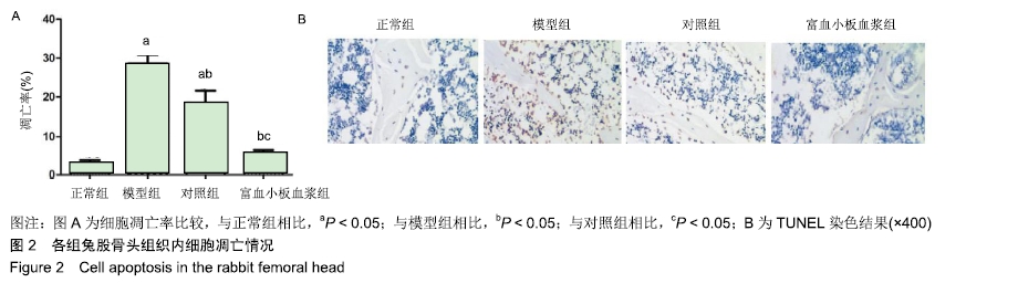
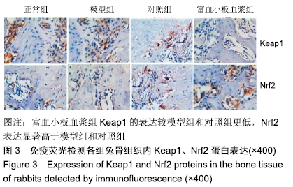
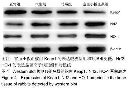
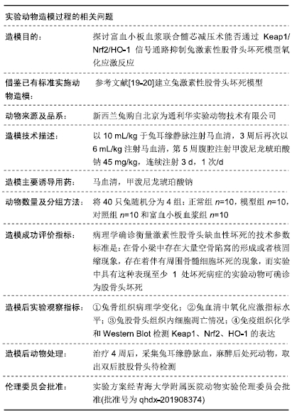
方法:将40只实验新西兰兔随机分为正常组、模型组、对照组和富血小板血浆组,每组10只。除正常组外其他3组兔在无菌环境下建立激素性股骨头坏死模型,术后4周向富血小板血浆组动物股骨头内髓芯减压后注射植入0.4 mL 3%的富血小板血浆,对照组兔只进行髓芯减压术治疗,对照组与模型组兔正常饲养。14周后苏木精-伊红染色观察各组兔股骨头骨髓腔内病理学变化和骨陷窝空缺率,检测各组兔血清中总抗氧化能力、超氧化物歧化酶、谷胱甘肽过氧化物酶、还原型谷胱甘肽及丙二醛等氧化应激指标活性,TUNEL检测股骨头组织内骨细胞凋亡情况,免疫荧光检测股骨头组织内Keap1、Nrf2分布,Western Blot检测股骨头组织内Keap1、Nrf2、HO-1蛋白表达。实验方案经青海大学附属医院动物实验伦理委员会批准(批准号为qhdx-201908374)。

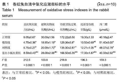
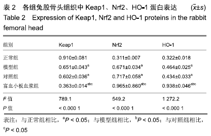
结果与结论:①与正常组相比,模型组骨组织内骨小梁变细,结构紊乱;对照组较模型组有所改善,骨小梁结构得到恢复,空骨陷窝减少(P < 0.05),富血小板血浆组经富血小板血浆联合髓芯减压治疗较对照组得到进一步改善,骨小梁结构更加完善,空骨陷窝进一步减少(P < 0.05),与正常组无显著差异(P > 0.05);②模型组血清中总抗氧化能力、超氧化物歧化酶、谷胱甘肽过氧化物酶和还原型谷胱甘肽的含量均显著低于正常组(P < 0.05),而丙二醛浓度显著高于正常组(P < 0.05);对照组以上指标稍有改善,但与模型组比较差异不显著(P > 0.05);富血小板血浆以上氧化应激指标较模型组和对照组明显改善(P < 0.05);③模型组股骨头组织内Keap1蛋白表达显著低于正常组(P < 0.05),Nrf2、HO-1蛋白表达显著高于正常组(P < 0.05);富血小板血浆组股骨头组织内Keap1的表达较模型组和对照组低(P < 0.05),Nrf2、HO-1的表达显著高于模型组和对照组(P < 0.05);④结果提示,富血小板血浆能有效抑制兔激素性股骨头坏死过程中氧化应激反应,该作用可能是通过激活Keap1/Nrf2/HO-1信号通路活性而发生的。

ORCID: 0000-0002-8371-2665(王哲)

中国组织工程研究杂志出版内容重点:组织构建;骨细胞;软骨细胞;细胞培养;成纤维细胞;血管内皮细胞;骨质疏松组织工程


关键词: 富血小板血浆, 股骨头坏死, 氧化应激, Keap1/Nrf2/HO-1信号通路

Abstract:

BACKGROUND: Oxidative stress plays an important role in femoral head necrosis. Platelet-rich plasma (PRP) contains growth factors that can accelerate fracture healing. PRP combined with core decompression can promote recovery from non-traumatic femoral head necrosis.

OBJECTIVE: To investigate whether PRP combined with core decompression can inhibit oxidative stress in steroid-induced avascular necrosis of the femoral head model via Keap1/Nrf2/HO-1 signaling pathway.

METHODS: Forty New Zealand rabbits were randomly divided into normal group, model group, control group and PRP group, with 10 rabbits in each group. In the model and PRP groups, a model of steroid-induced femoral head necrosis was established in a sterile environment. At 4 weeks after operation, the rabbits in the PRP group were injected with 0.4 mL of 3% PRP after core decompression. The control group received core decompression treatment, and the control and model groups were raised normally. After 14 weeks, the experimental animals were sacrificed. Hematoxylin-eosin staining was used to observe the pathological changes of bone marrow cavity and the vacancy rate of bone lacunae in the femoral head of each group. Total antioxidant capacity, superoxide dismutase, glutathione peroxidase, reduced glutathione, and malondialdehyde were detected. TUNEL was used to detect bone cell apoptosis in the femoral head. Immunofluorescence staining was used to determine the distribution of Keap1 and Nrf2. Western blot was used to measure Keap1, Nrf2, and HO-1 protein expression in the femoral head. Approval was obtained from the Animal Ethics Committee of the Affiliated Hospital of Qinghai University, approval No. qhdx-201908374.

RESULTS AND CONCLUSION: (1) Compared with the normal group, the trabecular bone in model group was thinned with structure disorder. Compared with the model group, the trabecular bone structure in control group was restored, and the number of vacant bone lacunae was reduced (P < 0.05). Compared with the control group, the animals treated with PRP combined with core decompression were further improved, the trabecular bone structure was further improved, and the number of vacant bone lacunae was further reduced (P < 0.05). Whereas there was no significant difference between the PRP group and normal group (P > 0.05). (2) The total antioxidant capacity and serum levels of superoxide dismutase, glutathione peroxidase, and reduced glutathione in the model group were significantly lower than those in normal animals (P < 0.05), while the MDA concentration was significantly higher than that in the normal group (P < 0.05). These oxidative stress indexes were slightly improved in the control group compared with the model group (P > 0.05), while these indexes were significantly improved in the PRP group than the model and control groups (P < 0.05). (3) The expression of Keap1 in the model group was significantly lower than that of the normal group   (P < 0.05), and the expression of Nrf2 and HO-1 protein was significantly higher than that of the normal group (P < 0.05). The expression of Keap1 in the PRP group was lower than that of the model and control groups (P < 0.05), and the expression of Nrf2 and HO-1 was significantly higher than that of the model and control groups (P < 0.05). Therefore, PRP can effectively inhibit oxidative stress in the process of steroid-induced femoral head necrosis, which may be caused by activating the Keap1/Nrf2/HO-1 signaling pathway.

Key words: platelet-rich plasma, femoral head necrosis, oxidative stress, Keap1/Nrf2/HO-1 signaling pathway

中图分类号: