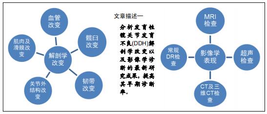
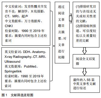
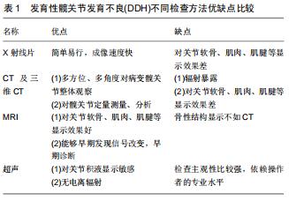
|
[1] JESSE MK, PETERSEN B, STRICKLAND C, et al. Normal anatomy and imaging of the hip: emphasis on impingement assessment. Semin Musculoskelet Radiol. 2013;17(3):229-247.
[2] SONG K, ANDERSON AE, WEISS JA, et al. Musculoskeletal models with generic and subject-specific geometry estimate different joint biomechanics in dysplastic hips. Comput Methods Biomech Biomed Engin. 2019;22(3):259-270.
[3] ONAY T, GUMUSTAS SA, CAGIRMAZ T, et al. Do the risk factors for developmental dysplasia of the hip differ according to gender? A look from another perspective. J Paediatr Child Health. 2019, 55(2):168-174.
[4] LERNER ZF, DEMERS MS, DELP SL, et al. How tibiofemoral alignment and contact locations affect predictions of medial and lateral tibiofemoral contact forces. J Biomech.2015;48(4):644.
[5] MÜLLER M, RAKOW A, WASSILEW GI, et al. Prediction of time to prosthesis implantation as a function of joint anatomy in patients with developmental dysplasia of the hip. J Orthop Surg Res. 2019;14(1):471.
[6] MINOKAWA S, NAITO M, KINOSHITA K, et al. Acetabular labrum blood flow in developmental dysplasia of the hip: an intraoperative in vivo study using laser Doppler flowmetry. J Orthop Surg Res. 2016;11(1):116.
[7] PETERSEN BD, WOLF B, LAMBERT JR, et al.Lateral acetabular labral length is inversely related to acetabular coverage as measured by lateral center edge angle of Wiberg. J Hip Preserv Surg.2016,3(3):190-196.
[8] HONG K, YUAN Z, LI J, et al. Femoral anteversion does not predict redislocation in children with hip dysplasia treated by closed reduction. Int Orthop. 2019;43(7):1635-1642.
[9] CHANG CY, HUANG AJ. MR Imaging of Normal Hip Anatomy. Magn Reson Imaging Clin N Am. 2013;21(1):1.
[10] 史立伟,赵群,张立军,等.血小板源生长因子A(PDGF-A)在发育性髋关节发育不良患儿圆韧带的分布和表达[J].中国矫形外科杂志,2010, 18(13):1098-1102.
[11] LEE SH, JANG WY, CHOI GW, et al. Is the Transverse Acetabular Ligament Hypertrophied and Hindering Reduction in Developmental Dysplasia of Hip?Arthroscopy. 2018;34(4): 1219-1226.
[12] LIU R, WEN X, TONG Z, et al.Changes of gluteus medius muscle in the adult patients with unilateral developmental dysplasia of the hip. BMC Musculoskelet Disord. 2012;13:101.
[13] 王亚捷,刘玲玲,孟燕,等.发育性髋脱位髋周肌肉的MR评价及T2-mapping定量分析[J].中华小儿外科杂志,2019,40(8):746-751.
[14] LE BOUTHILLIER A, RAKHRA KS, BELZILE EL, et al. Soft Tissue Structures Differ in Patients With Prearthritic Hip Disease. J Orthop Trauma. 2018;32(suppl):S30-S34.
[15] LI, WANG H, HE JY, et al. Inflammatory and fibrosis infiltration in synovium associated with the progression in developmental dysplasia of the hip. Mol Med Rep. 2019;19(4):2808-2816.
[16] DWEK JR. The hip: MR imaging of uniquely pediatric disorders. Magn Reson Imaging Clin N Am. 2009;17(3):509-520.
[17] HAEFELI PC, STEPPACHER SD, BABST D, et al. An increased iliocapsularis-to-rectus-femoris ratio is suggestive for instability in borderline hips. Clin Orthop Relat Res. 2015;473(12):3725-3734.
[18] ROSENBAUM DG, SERVAES S, BOGNER EA, et al. MR Imaging in Postreduction Assessment of Developmental Dysplasia of the Hip: Goals and Obstacles.Radiographics. 2016;36(3):840-854.
[19] KIM HT, KIM IB, LEE JS.MR-based parameters as a supplement to radiographs in managing developmental hip dysplasia.Clin Orthop Surg. 2011;3(3):202-210.
[20] DESAI AA, MARTUS JE, SCHOENECKER J, et al.Spica MRI after closed reduction for developmental dysplasia of the hip. Pediatr Radiol. 2011;41(4):525-529.
[21] JAMES S, MIOCEVIC M, MALARA F, et al. MR imaging findings of ace- tabular dysplasia in adults. Skeletal Radiol.2006;35(6): 378-384.
[22] LLOPIS E, CEREZAL L, KASSARJIAN A, et al. Direct MR arthrography of the hip with leg traction: feasibility for assessing articular cartilage. AJR.2008;190(4):1124-1128.
[23] TIDERIUS C, JARAMILLO D, CONNOLLY S, et al. Post - closed reduction per- fusion magnetic resonance imaging as a predictor of avascular necrosis in developmental hip dysplasia: a preliminary report. J Pediatr Orthop.2009;29(1):14-20.
[24] DOMAYER SE, MAMISCH TC, KRESS I, et al. Radial dGEMRIC in developmental dysplasia of the hip and in femoroacetabular impingement: preliminary results. Osteoarthritis Cartilage.2010; 18(11): 1421-1428
[25] LI LY, ZHANG LJ, LI QW, et al. Development of the osseous and cartilaginous acetabular index in normal children and those with develop- mental dysplasia of the hip: a cross-sectional study using MRI. J Bone Joint Surg Br.2012;94(12):1625-1631.
[26] FISHER R, O’BRIEN TS, DAVIS KM. Magnetic resonance imaging in congenital dysplasia of the hip. J Pediatr Orthop. 1991;11(5):617-622.
[27] DOUIRA-KHOMSI W, SMIDA M, LOUATI H, et al. Magnetic resonance evaluation of acetabular residual dysplasia in developmental dysplasia of the hip: a preliminary study of 27 patients. J Pediatr Orthop.2010;30(1):37-43.
[28] TAKUCHI R, KAMADA H, MISHIMA H, et al. Evaluation of the cartilaginous acetabulum by magnetic resonance imaging in developmental dysplasia of the hip. J Pediatr Orthop B.2014;23(3): 237-243.
[29] SHALABY MH, SAMIR S, DEIF A. CT measurement of femoral anteversion angle in patients with unilateral developmental hip dysplasia: A comparative study between 2D and 3D techniques. Egyptian Journal Of Radiology And Nuclear Medicine.2017; 48(3):639-643.
[30] CAI ZC, ZHAO Q, LI LY, et al. Can Computed Tomography Accurately Measure Acetabular Anterversion in Developmental Dysplasia of the Hip? Verification and Characterization Using 3D Printing Technology. J Pediatr Orthop.2018;38(4):e180-e185.
[31] WANG XY, PENG JP, ZHU JF, et al. Application of three-dimensional computerised tomography reconstruction and image processing technology in individual operation design of developmental dysplasia of the hip patients. Int Orthop.2016; 40(2):255-265.
[32] MASSA BSF, GUARNIERO R, GODOY RM JR, et al. Use of inlet radiographs in the assessment of reduction after the surgical treatment of developmental dysplasia of the hip. Bone Joint J. 2017; 99-B(5):697-701.
[33] MIMURA T, MORI K, KITAGAWA M, et al. Multiplanar evaluation of radiological findings associated with acetabular dysplasia and investigation of its prevalence in an Asian population: a CT-based study. BMC Musculoskelet Disord. 2017;18(1):50.
[34] YAMADA K, ENDO H, TETSUNAGA T, et al. Accuracy of Cup Positioning with the CT-Based 2D-3D Matched Navigation System: A Prospective, Randomized and Controlled Study. J Arthroplasty.2018;33(1):136-143.
[35] GRISSOM L, HARCKE HT, THACKER M. Imaging in the surgical manage
ment of developmental dislocation of the hip. Clin Orthop.2008;466(4):791-801.
[36] IA J, LI L, ZHANG L, et al. Three dimensional - CT evaluation of fem- oral neck anteversion,acetabular anteversion and combined antever- sion in unilateral DDH in an early walking age group. Int Orthop.2012;36(1):119-124.
[37] ZHANG J, WANG L, MAO Y, et al. The use of combined anteversion in total hip arthroplasty for patients with developmental dysplasia of the hip. J Arthroplasty.2014;29(3):621-625.
[38] LANGLOTZ F, BACHLER R, BERLEMANN U, et al. Computer assistance for pelvic osteotomies. Clin Orthop.1998;354:92-102.
[39] VAN BOSSE HJ, LEE D, HENDERSON ER, et al. Pelvic positioning creates error in CT acetabular measurements. Clin Orthop.2011;469(6):1683-16891.
[40] FRICK SL, KIM SS, WENGER DR. Pre- and postoperative three-dimensional computed tomography analysis of triple innominate osteotomy for hip dysplasia. J Pediatr Orthop. 2000; 20(1):116-123.
[41] ZHAO X, CHOSA E, TOTORIBE K, et al. Effect of periacetabular osteotomy for acetabular dysplasia clarified by three-dimensional finite element analysis. J Orthop Sci.2010;15(5):632-640.
[42] NIE Y, PEI FX, SHEN B, et al. Implication of Acetabular Width on the Anteroposterior Pelvic Radiograph of Patients With Developmental Dysplasia of the Hip. J Arthroplasty.2015;30(3): 489-494.
[43] ISMIARTO YD, AGRADI P, HELMI ZN.Comparison of Interobserver Reliability between Junior and Senior Resident in Assessment of Developmental Dysplasia of The Hip Severity using Tonnis and International Hip Dysplasia Institute Radiological Classification.Malays Orthop J. 2019;13(3):60-65.
[44] AIUM-ACR-SPR-SRU practice for the performance of an ultrosound examination for detection and assessment of developmental of the hip. J Ultrosoud Med.2018;37(11):E1-E5.
[45] D’AlESSANDRO M, DOW K. Investigating the need for routine ultrasound screening to detect developmental dysplasia of the hip in infants born with breech presentation. Paediatr Child Health. 2019;24(2):e88-e93.
[46] SPAANS AJ, BEEK FJA, UITERWAAL CSPM, et al.Correlation between ultrasonic and radiographic imaging of developmental dysplasia of the hip. J Child Orthop.2019;13(2):155-160.
[47] PARK HK, KANG EY, LEE SH, et al. The Utility of Ultrasonography for the Diagnosis of Developmental Dysplasia of Hip Joint in Congenital Muscular Torticollis. Ann Rehabil Med. 2013;37(1):26-32.
[48] HUANG GL., CHEN YQ. Role of ultrasonography in the diagnosis of developmental dysplasia of the hip. Chinese Journal of Medical Imaging Technology.2009,25(4):712-714.
[49] ALEXIEV VA, HARCKE HT, KUMAR SJ. Residual dysplasia after successful Pavlik harness treatment: early ultrasound predictors. J Pediatr Orthop. 2006,26(1):16 - 23.
[50] TRÉGUIER C, CHAPUIS M, BRANGER B, et al. Pubo-femoral distance: an easy sonographic screening test to avoid late diagnosis of developmental dysplasia of the hip. Eur Radiol. 2013; 23(3):836-844.
[51] ARTI H, MEHDINASAB SA, ARTI S. Comparing results of clinical versus ultrasonographic examination in developmental dysplasia of hip. J Res Med Sci. 2013;18(12):1051-1055.
[52] PUHAN MA, WOOLACOTT N, KLEIJNEN J, et al. Observational studies on ultrasound screening for developmental dysplasia of the hip in newborns a systematic review. Ultraschall Med. 2003; 24(6):377-382.
[53] OMEROGLU H, CAYLAK R, INAN U, et al. Ultrasonographic Graf type IIa hip needs more consideration in newborn girls. J Child Orthop.2013;7(2):95-98.
[54] TSCHAUNER C, FÜRNTRATH F, SABA Y, et al. Developmental dysplasia of the hip: impact of sonographic newborn hip screening on the outcome of early treated decentered hip joints - a single center retrospective comparative cohort study based on Graf’s method of hip ultrasonography. J Child Orthop. 2011;5(6): 415-424.
[55] FARR S, GRILL F, MÜLLER D. When is the optimal time for hip ultrasound screening. Orthopade. 2008;37(6):532,534-536, 538-540.
|