中国组织工程研究 ›› 2020, Vol. 24 ›› Issue (10): 1502-1507.doi: 10.3969/j.issn.2095-4344.2217

• 组织工程骨及软骨材料 tissue-engineered bone and cartilage materials • 上一篇    下一篇

骨水泥间隔器表面微观形貌改变对诱导膜内成骨因子表达的影响

李树源1,周琦石2,李  悦2,林知毅3,周宏亮1,申  震1,胡  柽1,杨佳宝1   

  1. 1广州中医药大学第一临床医学院,广东省广州市  510405;2广州中医药大学第一附属医院一骨科,广东省广州市  510405;3广州市番禺区中医院,广东省广州市  511400
  • 收稿日期:2019-06-03 修回日期:2019-06-11 接受日期:2019-07-10 出版日期:2020-04-08 发布日期:2020-02-14
  • 通讯作者: 周琦石,副教授,广州中医药大学第一附属医院一骨科,广东省广州市 510405
  • 作者简介:李树源,男,1991年生,山西省汾阳市人,汉族,广州中医药大学第一临床医学院在读硕士,主要从事创伤骨科研究。
  • 基金资助:
    国家自然科学基金资助项目(81674001)

Effects of changes in surface morphology of bone cement spacer on the expression of bone growth factor in induced membrane 

Li Shuyuan1, Zhou Qishi2, Li Yue2, Lin Zhiyi3, Zhou Hongliang1, Shen Zhen1, Hu Cheng1, Yang Jiabao1   

  1. 1The First Clinical Medical College, Guangzhou University of Chinese Medicine, Guangzhou 510405, Guangdong Province, China; 2First Department of Orthopedics, First Affiliated Hospital of Guangzhou University of Chinese Medicine, Guangzhou 510405, Guangdong Province, China; 3Panyu Hospital of Chinese Medicine, Guangzhou 511400, Guangdong Province, China
  • Received:2019-06-03 Revised:2019-06-11 Accepted:2019-07-10 Online:2020-04-08 Published:2020-02-14
  • Contact: Zhou Qishi, Associate professor, First Department of Orthopedics, First Affiliated Hospital of Guangzhou University of Chinese Medicine, Guangzhou 510405, Guangdong Province, China
  • About author:Li Shuyuan, Master candidate, The First Clinical Medical College, Guangzhou University of Chinese Medicine, Guangzhou 510405, Guangdong Province, China
  • Supported by:
     the National Natural Science Foundation of China, No. 81674001

摘要:

文题释义:
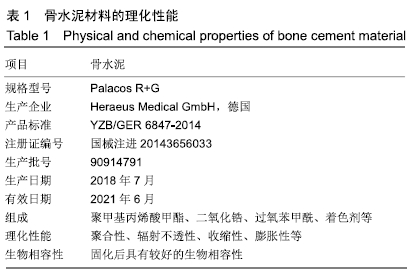
聚甲基丙烯酸甲酯骨水泥:为临床上常用的骨科植入材料,具有很大的抗压能力,抗压缩能力为97 MPa,也具有良好的生物相容性。1987年Galibert等首次报道用聚甲基丙烯酸甲酯骨水泥来治疗椎体血管瘤,并将此技术称之为经皮椎体成形。1970年Buchholz首次应用载抗生素骨水泥控制关节感染,目前以聚甲基丙烯酸甲酯骨水泥作为抗生素的缓释载体被广泛应用于人工全髋关节置换及骨髓炎的治疗。
转化生长因子β1:是具有多种功能的蛋白多肽,是“转化生长因子β超家族”成员之一,大量存在于骨组织与血小板中,可以刺激间充质干细胞的增殖、分化,并抑制间充质干细胞向脂肪细胞分化,也促进成骨细胞、成软骨细胞的增殖及细胞外基质的合成,诱导膜内成骨和软骨内成骨。


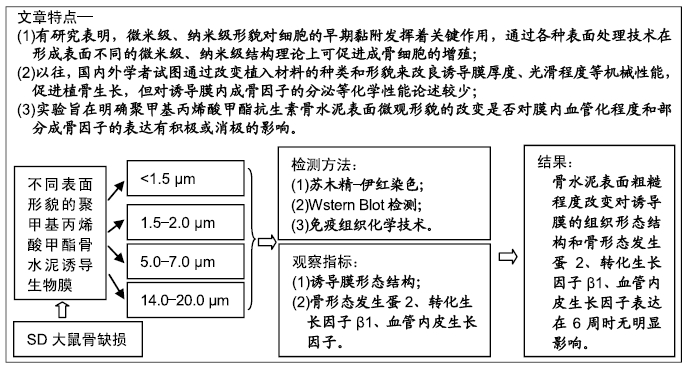
背景:目前国内外学者试图通过改变植入材料的种类和形貌、改良诱导膜厚度、光滑程度等机械化学性能来促进植骨生长。

目的:比较大鼠股骨骨缺损处不同表面粗糙程度聚甲基丙烯酸甲酯骨水泥形成的诱导膜在膜内血管化程度和部分成骨因子表达的差异。

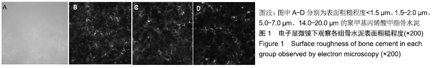
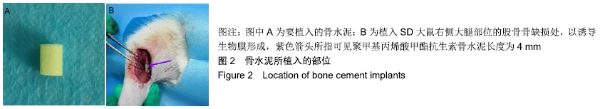
方法:取48只雄性SD大鼠(购自广州中医药大学实验动物中心)建立大鼠临界尺寸股骨缺损模型,按随机数字表法分为A、B、C、D组,分别在股骨骨缺损处植入表面粗糙度<1.5 µm、1.5-2.0 µm、5.0-7.0 µm、14.0-20.0 µm的聚甲基丙烯酸甲酯骨水泥占位器。植入6周大鼠体内诱导膜形成后取出骨水泥周围的诱导膜,苏木精-伊红染色观察诱导膜病理组织形态结构变化,采用Western Blot印迹方法和免疫组织化学染色法对诱导膜中骨形态发生蛋白2、转化生长因子β1、血管内皮生长因子蛋白进行定量和定性分析。实验获得广州中医药大学动物实验伦理委员会批准,批准号:20181101006。

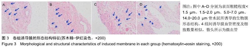
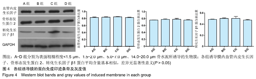
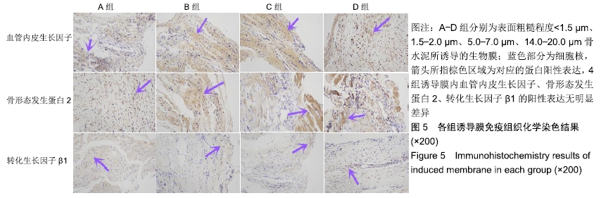
结果与结论:①苏木精-伊红染色显示,4种表面粗糙程度不同的骨水泥均可以形成较为规则的诱导膜,4组诱导膜之间血管化程度和细胞的数量大体相似;②Western Blot印迹检测显示,各组诱导膜内骨形态发生蛋2、转化生长因子β1、血管内皮生长因子蛋白平均含量基本相似(P > 0.05);③免疫组织化学染色显示,各组诱导膜内骨形态发生蛋2、转化生长因子β1、血管内皮生长因子蛋白阳性表达基本相似(P > 0.05);④结果表明,骨水泥表面粗糙程度改变对诱导膜的组织形态结构和骨形态发生蛋2、转化生长因子β1、血管内皮生长因子表达在6周时无明显影响。

ORCID: 0000-0003-1405-0765(李树源)

中国组织工程研究杂志出版内容重点:生物材料;骨生物材料; 口腔生物材料; 纳米材料; 缓释材料; 材料相容性;组织工程

关键词: 聚甲基丙烯酸甲酯骨水泥, 表面形貌, 诱导膜, 微血管, 骨生长因子, 骨形态发生蛋2, 转化生长因子β1, 血管内皮生长因子

Abstract:

BACKGROUND: At present, scholars at home and abroad try to promote the growth of bone graft by changing the types and morphology of implant materials, and improving the thickness and smoothness of induced membrane.

OBJECTIVE: To compare the degree of vascularization and the expression of some osteogenic factors in the polymethylmethacrylate bone cement membrane with different surface roughness degrees at different femoral bone defects in rats.

METHODS: Forty-eight male Sprague-Dawley rats (purchased from the Experimental Animal Center of Guangzhou University of Chinese Medicine) were used to establish rat models of critical-size femur defect. These rat modes were then randomly divided into groups A, B, C and D. Polymethylmethacrylate bone cement occupiers with surface roughness < 1.5 μm, 1.5-2.0 μm, 5.0-7.0 μm, and 14.0-20.0 μm were implanted in the femoral bone defects, respectively. At 6 weeks after implantation of bone cement spacer, the induced membranes around bone cement were removed. The morphological changes of the induced membranes were observed by hematoxylin-eosin staining. Western blotting and immunohistochemical staining were used to detect bone morphogenetic protein 2, transforming growth factor beta 1 and vascular endothelial growth factor protein in the induced membranes for quantitative and qualitative analyses. This study was approved by the Animal Ethics Committee of Guangzhou University of Chinese Medicine, China (approval No. 20181101006).

RESULTS AND CONCLUSION: Hematoxylin-eosin staining showed that four kinds of bone cement with different surface roughness degrees could form a relatively regular induced membrane. The degree of vascularization and the number of cells in the induced membrane were similar among the four groups. Western blot analysis showed that the average levels of bone morphogenetic protein 2, transforming growth factor β1 and vascular endothelial growth factor protein were similar among four groups (P > 0.05). Immunohistochemical staining revealed that the positive expression of bone morphogenetic protein 2, transforming growth factor β1 and vascular endothelial growth factor protein was similar among four groups (P > 0.05). These findings suggest that the change of surface roughness of bone cement has no significant effect on the histomorphological structure of induced membrane and the expression of bone morphogenetic protein 2, transforming growth factor β1 and vascular endothelial growth factor in induced membrane at 6 weeks after implantation of bone cement spacer.

Key words: polymethylmethacrylate bone cement, surface morphology, induced membrane, microvessel, bone growth factor, bone morphogenetic protein 2, transforming growth factor β1, vascular endothelial growth factor

中图分类号: