中国组织工程研究 ›› 2020, Vol. 24 ›› Issue (29): 4632-4637.doi: 10.3969/j.issn.2095-4344.2812
• 皮肤粘膜组织构建 skin and mucosal tissue construction • 上一篇 下一篇
L-精氨酸通过L-Arg-NO途径促进大鼠背部跨区皮瓣的成活
李文波1,2,石 杰1,2,时培晟1,薛 云1,黄 强1,李闯兵1,高秋明1
- 1解放军联勤保障部队第九四〇医院创伤骨科,甘肃省兰州市 730050;2甘肃中医药大学,甘肃省兰州市 730000
L-arginine promotes the survival of extended dorsal perforator skin flap through activating L-arginine-nitric oxide pathway
Li Wenbo1, 2, Shi Jie1, 2, Shi Peisheng1, Xue Yun1, Huang Qiang1, Li Chuangbing1, Gao Qiuming1
- 1Department of Traumatic Orthopedics, the 940th Hospital of the PLA Joint Logistic Support Force, Lanzhou 730050, Gansu Province, China; 2Guansu University of Chinese Medicine, Lanzhou 730000, Gansu Province, China
摘要:

文题释义:
跨区皮瓣:穿支血管体区是某一源动脉呈三维立体网状结构所供应的所有解剖区域,是设计皮瓣的解剖学基础。临床上大面积皮肤软组织缺损时常需扩大切取皮瓣,皮瓣内包含2个以上穿支血管体区,称为跨区皮瓣。
Choke区:穿支血管呈树状分支,且口径逐渐减小,并与周围临近穿支血管形成血管网状连接,及“choke vessels”,两相邻穿支血管之间的区域称为Choke区。某一穿支血管不能通过choke区无限制向皮瓣远端供血,穿支皮瓣的缺血、坏死经常出现在choke区域,choke vessels成了穿支皮瓣血运的最大阻力。
背景:有研究表明一氧化氮可有效改善皮瓣术后血供,促进皮瓣成活,但具体机制尚不清楚。
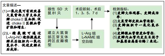
目的:通过实验验证L-Arg-NO途径在L-精氨酸促进大鼠背部跨区皮瓣成活中的作用。
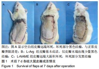
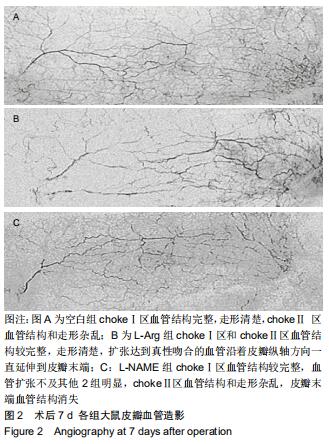
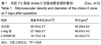
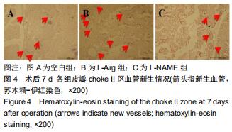
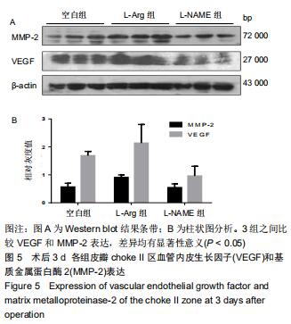
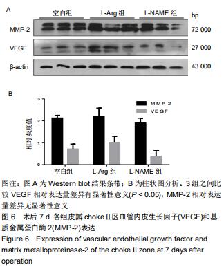
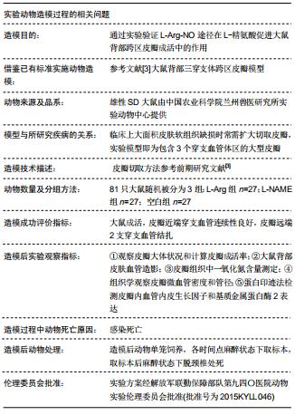
方法:成功建立大鼠背部三穿支体跨区皮瓣模型的雄性SD大鼠81只,随机被分为3组,分别于术后即刻、术后1-7 d腹腔注射不同药物,L-Arg组注射L-精氨酸 400 mg/(kg·d);L-NAME组注射一氧化氮合酶抑制剂40 mg/(kg·d);空白组注射等体积等渗氯化钠溶液。术后7 d观察皮瓣成活情况,计算皮瓣成活率;颈总动脉灌注明胶-氧化铅行血管造影,观察血管走形和分布;硝酸还原酶法测定术后即刻、1,3,5,7 d chokeⅡ区一氧化氮水平;苏木精-伊红染色观察choke Ⅱ区新生血管密度和管径;蛋白印记法检测术后3,7 d chokeⅡ区血管内皮生长因子和基质金属蛋白酶2蛋白表达。实验方案经解放军联勤保障部队第九四〇医院动物实验伦理委员会批准(批准号为2015KYLL046)。
结果与结论:①术后7 d,L-Arg组大鼠皮瓣成活率最高(89.47±3.17)%,3组比较差异有显著性意义(F=49.908,P < 0.001);②术后7 d,L-Arg组choke Ⅱ区血管结构较完整,走形清楚,扩张达到真性吻合的血管沿着皮瓣纵轴方向一直延伸到皮瓣末端,空白组和L-NAME组chokeⅡ区血管结构和走形杂乱,L-NAME组皮瓣末端血管结构消失;③L-Arg组一氧化氮水平术后开始升高,术后3 d达到高峰,之后逐渐下降;④术后7 d,L-Arg组新生血管密度和管径最高,3组比较差异有显著性意义(均P < 0.001);⑤术后3 d,L-Arg组血管内皮生长因子和基质金属蛋白酶2蛋白表达量最高,3组比较差异有显著性意义(均P < 0.05);术后7 d,L-Arg组血管内皮生长因子和基质金属蛋白酶2蛋白表达量最高,3组之间比较血管内皮生长因子差异有显著性意义(P < 0.05),基质金属蛋白酶2蛋白差异无显著性意义;⑥结果说明,一氧化氮可有效改善大鼠背部三穿支体皮瓣chokeⅡ区血供,促进chokeⅡ区微血管扩张和增生,而L-精氨酸可通过L-Arg-NO途径提高皮瓣术后组织内一氧化氮水平,促进皮瓣术后成活。
ORCID: 0000-0002-1812-9457(李文波);0000-0003-2403-1082(高秋明)中国组织工程研究杂志出版内容重点:组织构建;骨细胞;软骨细胞;细胞培养;成纤维细胞;血管内皮细胞;骨质疏松;组织工程
中图分类号: