中国组织工程研究 ›› 2021, Vol. 25 ›› Issue (25): 3988-3993.doi: 10.12307/2021.009

• 干细胞移植 stem cell transplantation • 上一篇    下一篇

过表达脯氨酰寡肽酶的骨髓间充质干细胞修复肝纤维化模型大鼠

郝晓娜,张英杰,李玉云,许  涛   

  1. 蚌埠医学院检验医学院,安徽省蚌埠市  233030
  • 收稿日期:2020-09-17 修回日期:2020-09-19 接受日期:2020-10-29 出版日期:2021-09-08 发布日期:2021-03-25
  • 通讯作者: 张英杰,硕士,副教授,蚌埠医学院检验医学院,安徽省蚌埠市 233030
  • 作者简介:郝晓娜,女,1981年生,河北省邯郸市人,汉族,硕士,讲师,主要从事间充质干细胞研究。
  • 基金资助:
    蚌埠医学院科技发展基金(BYKF1729),项目负责人:张英杰;安徽省教育厅高校科学研究重点项目(KJ2019A0391),项目负责人:张英杰

Bone marrow mesenchymal stem cells overexpressing prolyl oligopeptidase on the repair of liver fibrosis in rat models

Hao Xiaona, Zhang Yingjie, Li Yuyun, Xu Tao   

  1. School of Laboratory Medicine, Bengbu Medical College, Bengbu 233030, Anhui Province, China
  • Received:2020-09-17 Revised:2020-09-19 Accepted:2020-10-29 Online:2021-09-08 Published:2021-03-25
  • Contact: Zhang Yingjie, Master, Associate professor, School of Laboratory Medicine, Bengbu Medical College, Bengbu 233030, Anhui Province, China
  • About author:Hao Xiaona, Master, Lecturer, School of Laboratory Medicine, Bengbu Medical College, Bengbu 233030, Anhui Province, China
  • Supported by:
    the Science and Technology Development Fund of Bengbu Medical College, No. BYKF1729 (to ZYJ); the Key Scientific Research Project of Colleges and Universities of Anhui Provincial Department of Education, No. KJ2019A0391 (to ZYJ)

摘要:

文题释义:
脯氨酰寡肽酶:一种丝氨酸蛋白酶,由约700个氨基酸残基组成,分子质量为 70-80 ku,与其他组织相比,脯氨酰寡肽酶在肝脏的活性最高,脯氨酰寡肽酶均衡地分布于肝细胞、库普弗细胞的胞质和胞核内,而肝细胞与库普弗细胞均是肝损伤修复的关键细胞。
肝星状细胞:肝星状细胞占肝脏固有细胞总数的15%,它是纤维化肝组织细胞外基质的主要来源,肝星状细胞的大量活化、增殖是肝纤维化发生、发展的重要环节。

背景:如何能使移植后的间充质干细胞更好地存活并发挥其功能是目前间充质干细胞研究的热点之一。
目的:探索过表达脯氨酰寡肽酶的骨髓间充质干细胞对大鼠肝纤维化的修复作用,并探讨相关机制。
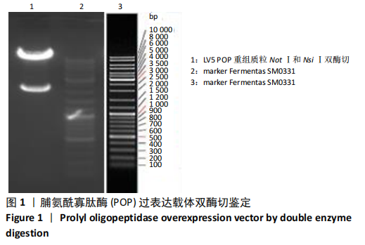
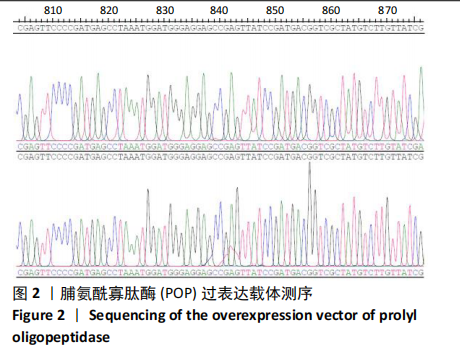
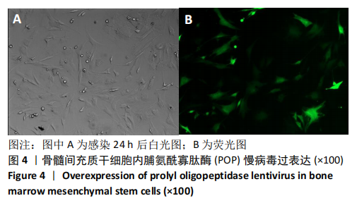
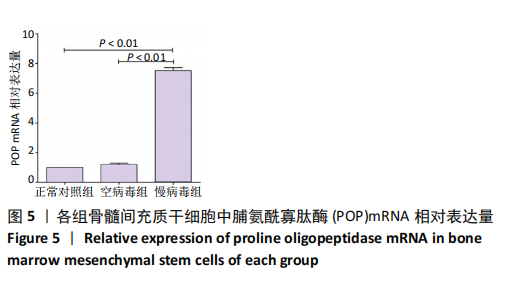
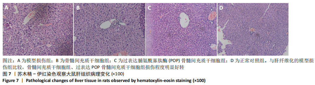
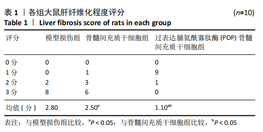
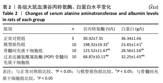
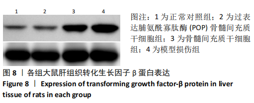
方法:35只雄性SD大鼠随机选取5只进入正常对照组(n=5),其余30只大鼠采用皮下多点注射四氯化碳花生油溶液方法制备大鼠肝纤维化模型,造模成功后被随机分为模型损伤组(n=10)、骨髓间充质干细胞组(n=10)、过表达脯氨酰寡肽酶的骨髓间充质干细胞组(n=10),经尾静脉分别注射生理盐水、1×10⁶骨髓间充质干细胞、1×10⁶过表达脯氨酰寡肽酶的骨髓间充质干细胞。3周后将大鼠处死,收集各组大鼠下腔静脉血,检测肝功能、肝纤维化指标;摘取肝脏行苏木精-伊红染色观察病理变化,荧光显微镜下观察细胞定位情况,Western blot 法检测肝组织转化生长因子β蛋白的表达水平。
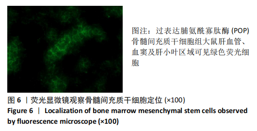
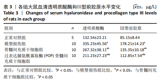
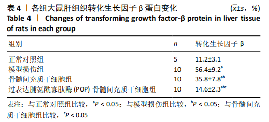
结果与结论:①与模型损伤组比较,2个细胞移植组大鼠肝功能、肝纤维化指标均明显改善,且过表达脯氨酰寡肽酶的骨髓间充质干细胞组改善更明显(P < 0.05);②大鼠肝组织冰冻切片荧光显微镜下观察显示:过表达脯氨酰寡肽酶的骨髓间充质干细胞组可见绿色荧光细胞,说明骨髓间充质干细胞成功在大鼠肝脏定植;③肝组织苏木精-伊红染色显示,2个细胞移植组肝纤维化程度明显改善,且过表达脯氨酰寡肽酶的骨髓间充质干细胞组肝脏病理更接近于正常;④2个细胞移植组转化生长因子β蛋白表达较模型损伤组均有明显降低(P < 0.05),且过表达脯氨酰寡肽酶的骨髓间充质干细胞组降低更明显(P < 0.05);⑤结果表明,过表达脯氨酰寡肽酶能够显著提高骨髓间充质干细胞对肝纤维化的修复能力。
https://orcid.org/0000-0001-8819-2219(张英杰) 

中国组织工程研究杂志出版内容重点:干细胞;骨髓干细胞;造血干细胞;脂肪干细胞;肿瘤干细胞;胚胎干细胞;脐带脐血干细胞;干细胞诱导;干细胞分化;组织工程

关键词: 干细胞, 骨髓间充质干细胞, 脯氨酰寡肽酶, 过表达, 肝纤维化, 转化生长因子β, 大鼠

Abstract: BACKGROUND: How to make the transplanted mesenchymal stem cells survive and function better is one of the hot spots in the research of mesenchymal stem cells.
OBJECTIVE: To investigate the repair function of bone marrow mesenchymal stem cells overexpressing prolyl oligopeptidase on liver fibrosis in rats and explore the related mechanisms.
METHODS:  Five rats were randomly selected from 35 male Sprague-Dawley rats into the normal control group (n=5). The remaining 30 rats were injected subcutaneously with CCl4 peanut oil solution to prepare rat liver fibrosis models. After successful modeling, they were randomly divided into model injury group (n=10), bone marrow mesenchymal stem cells group (n=10), and prolyl oligopeptidase-overexpressed bone marrow mesenchymal stem cells group (n=10). The model injury group was given normal saline via the tail vein. Bone marrow mesenchymal stem cells group was given 1×10⁶ bone marrow mesenchymal stem cells. The prolyl oligopeptidase overexpression bone marrow mesenchymal stem cells group was given 1×10⁶ prolyl oligopeptidase-overexpressed bone marrow mesenchymal stem cells. Three weeks later, the rats were sacrificed. The inferior vena cava blood of each group was collected to detect liver function and liver fibrosis indicators. The liver was taken out and stained with hematoxylin-eosin to observe the pathological changes. Cell localization was observed under a fluorescence microscope. Western blot assay was conducted to detect the expression level of transforming growth factor-β protein in the liver.  
RESULTS AND CONCLUSION: (1) The liver function and fibrosis indexes of rats in the bone marrow mesenchymal stem cells group and prolyl oligopeptidase-overexpressed bone marrow mesenchymal stem cells group were significantly improved compared with the model injury group, and the improvement was more obvious in the prolyl oligopeptidase-overexpressed bone marrow mesenchymal stem cells group (P < 0.05). (2) Fluorescence microscope of frozen section of rat liver tissue showed that there were green fluorescent cells in the prolyl oligopeptidase-overexpressed bone marrow mesenchymal stem cells group, indicating that bone marrow mesenchymal stem cells were successfully colonized in the rat liver. (3) Hematoxylin-eosin staining of liver tissue indicated that the degree of liver fibrosis in the bone marrow mesenchymal stem cells and prolyl oligopeptidase-overexpressed bone marrow mesenchymal stem cells groups was significantly improved, and the liver pathology of the prolyl oligopeptidase-overexpressed bone marrow mesenchymal stem cells group was closer to normal. (4) The expression level of transforming growth factor-β protein in the bone marrow mesenchymal stem cells group and prolyl oligopeptidase-overexpressed bone marrow mesenchymal stem cells group was significantly lower than that of the model injury group (P < 0.05), and the decrease in the prolyl oligopeptidase-overexpressed bone marrow mesenchymal stem cells group was more obvious (P < 0.05). (5) Results verify that overexpression of prolyl oligopeptidase can significantly improve the repair ability of bone marrow mesenchymal stem cells on liver fibrosis.

Key words: stem cells, bone marrow mesenchymal stem cells, prolyl oligopeptidase, overexpression, liver fibrosis, transforming growth factor-β, rats

中图分类号: