中国组织工程研究 ›› 2020, Vol. 24 ›› Issue (29): 4680-4685.doi: 10.3969/j.issn.2095-4344.2780
• 组织构建实验造模 experimental modeling in tissue construction • 上一篇 下一篇
全身应用神经生长因子对大鼠胫骨干骨折早期愈合作用及骨形态发生蛋白2和血管内皮生长因子表达的影响
刘国铭1,2,王钦奋2,林克凤2,周仕国2,陈祖星3,林世水2
1福建医科大学省立临床学院,福建省福州市 350001;2福建省立金山医院骨科,福建省福州市 350001;3福建省立医院骨科,福建省福州市 350001
Whole body application of nerve growth factor promotes early healing of tibial shaft fracture and improves expression of bone morphogenetic protein-2 and vascular endothelial growth factor in rats
Liu Guoming1, 2, Wang Qinfen2, Lin Kefeng2, Zhou Shiguo2, Chen Zuxing3, Lin Shishui2
1Provincial Clinical College of Fujian Medical University, Fuzhou 350001, Fujian Province, China; 2Department of Orthopaedics, Jinshan Hospital of Fujian Provincial Hospital, Fuzhou 350001, Fujian Province, China; 3Department of Orthopaedics, Fujian Provincial Hospital, Fuzhou 350001, Fujian Province, China
摘要:
文题释义:
神经生长因子:神经生长因子家族包括神经生长因子、脑源性神经营养因子、神经营养素3、神经营养素4/5、神经营养素6和神经营养素7等,主要为前4种。神经生长因子为神经生长因子家族中最主要的一类,在组织中主要以前体的形式存在,在颌下腺中加工形成成熟的神经生长因子,能促进中枢和外周神经元的生长、发育、分化、成熟,维持神经系统的正常功能,加快神经系统损伤后的修复。神经生长因子广泛分布于机体各组织器官中(包括脑),在靶组织中的浓度与交感神经和感觉神经在靶区分支的密度和mRNA的含量有关。
骨折愈合:是一个复杂而连续的过程,从组织学和细胞学的变化,通常将其分为3个阶段,但三者之间又不可截然分开,而是互相交织逐渐演进,包括血肿炎症机化期、原始骨痂形成期、骨板形成塑形期。
背景:骨折愈合是一种十分复杂的多阶段生理过程,由多种细胞协调参与。有关神经因素对于骨折愈合过程的影响逐渐被重视和深入研究,神经生长因子是一种能够促进细胞生长的生物活性复合蛋白,其促进骨折愈合过程中所发挥的作用机制尚不明确。
目的:观察神经生长因子对大鼠胫骨骨折愈合的促进作用及愈合过程中机制。
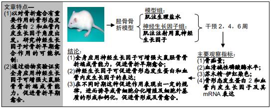
方法:将72只雄性SD大鼠随机分为模型组和神经生长因子组,构建胫骨干闭合骨折模型并应用髓内固定。神经生长因子组给予肌肉注射,注射用鼠神经生长因子;模型组给予肌肉注射生理盐水。实验于2018-05-30经福建医科大学实验动物伦理委员会批准,批准号:FJYKDX-2018-035。
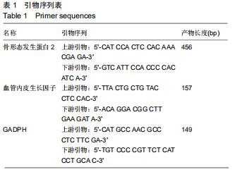
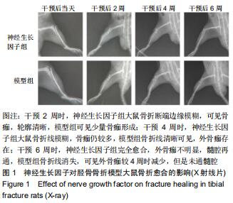
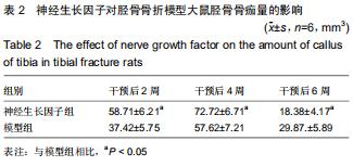
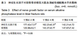
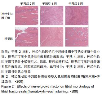
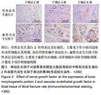
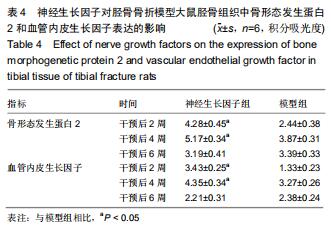
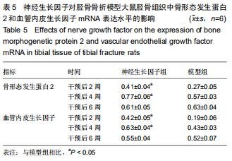
结果与结论:①干预2周和4周时,神经生长因子组骨痂体积显著高于模型组,干预6周时,神经生长因子组骨折线消失,髓腔再通,骨折早于模型组达到完全愈合;②苏木精-伊红染色显示干预2周和4周时,神经生长因子组骨小梁生长致密,可见大量成骨细胞生长;③干预2周和4周时,神经生长因子组血清碱性磷酸酶含量明显高于模型组;④免疫组化结果显示,干预2周和4周时,神经生长因子组骨痂内骨形态发生蛋白2和血管内皮生长因子阳性表达均显著多于模型组;⑤定量PCR结果显示,干预2周和4周时,神经生长因子组骨形态发生蛋白2和血管内皮生长因子 mRNA表达水平均高于模型组;⑥提示全身应用神经生长因子可增强大鼠胫骨骨折端成骨能力,促进骨折早期愈合;同时神经生长因子可促进骨形态发生蛋白2和血管内皮生长因子的表达,并在不同时期这种促进作用表现出一定的规律,进而诱导成骨细胞分化增殖及细胞外基质的形成和钙化,促进骨形成及骨愈合。
ORCID: 0000-0002-8369-5711(刘国铭)中国组织工程研究杂志出版内容重点:组织构建;骨细胞;软骨细胞;细胞培养;成纤维细胞;血管内皮细胞;骨质疏松;组织工程
中图分类号: