中国组织工程研究 ›› 2021, Vol. 25 ›› Issue (5): 754-760.doi: 10.3969/j.issn.2095-4344.3011

• 组织构建细胞学实验 cytology experiments in tissue construction • 上一篇    下一篇

转化生长因子β1诱导心肌成纤维细胞转分化过程中Hedgehog信号通路的作用机制

聂慧娟,黄织春   

  1. 内蒙古医科大学附属医院心血管内科,内蒙古自治区呼和浩特市   010059
  • 收稿日期:2020-05-19 修回日期:2020-05-21 接受日期:2020-06-12 出版日期:2021-02-18 发布日期:2020-12-01
  • 通讯作者: 黄织春,博士,主任医师,教授,内蒙古医科大学附属医院心血管内科,内蒙古自治区呼和浩特市 010059
  • 作者简介:聂慧娟,1983年生,内蒙古自治区呼和浩特市人,2018年重庆医科大学毕业,博士,副主任医师,副教授,主要从事冠心病的基础研究与临床诊疗研究。
  • 基金资助:
    内蒙古自然科学基金面上项目(2016MS(LH)0802)

The role of Hedgehog signaling pathway in transforming growth factor beta1-induced myofibroblast transdifferentiation

Nie Huijuan, Huang Zhichun   

  1. Department of Cardiology, Affiliated Hospital of Inner Mongolia Medical University, Hohhot 010059, Inner Mongolia Autonomous Region, China
  • Received:2020-05-19 Revised:2020-05-21 Accepted:2020-06-12 Online:2021-02-18 Published:2020-12-01
  • Contact: Huang Zhichun, MD, Chief physician, Professor, Department of Cardiology, Affiliated Hospital of Inner Mongolia Medical University, Hohhot 010059, Inner Mongolia Autonomous Region, China
  • About author:Nie Huijuan, MD, Associate chief physician, Associate professor, Department of Cardiology, Affiliated Hospital of Inner Mongolia Medical University, Hohhot 010059, Inner Mongolia Autonomous Region, China
  • Supported by:
    the Natural Science Foundation of Inner Mongolia Autonomous Region (General Project), No. 2016MS(LH)0802

摘要:

文题释义:
上皮间质转分化:是人体正常发育过程中或者机体在各种致病因素刺激下,上皮表型向间质表型转分化的过程,进而失去正常功能,组织器官发生纤维化异常增殖。
Hedgehog信号通路:是与人体生长、发育及疾病的发生发展有着密切关系的细胞通路,参与胚胎发育、器官形成及多种疾病损伤发病过程,但在心肌纤维化中的相关作用不明确,此次研究探讨其参与心肌纤维化的作用机制。

背景:近年来越来越多的研究表明Hedghog信号通路异常活化广泛参与全身多组织器官疾病的损伤修复过程,但其在心肌纤维化中的相关作用尚不明确。
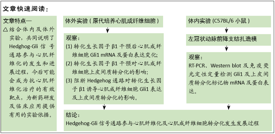
目的:研究阻断Hedgehog-Gli信号通路对转化生长因子β1诱导的心肌成纤维细胞上皮间质转分化过程的影响。
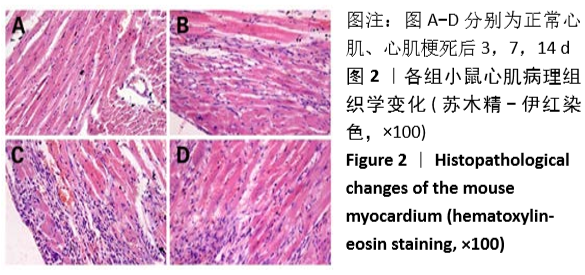
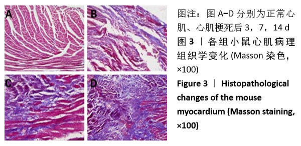
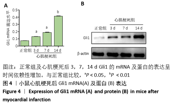
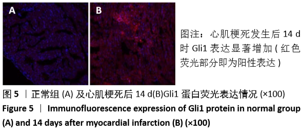
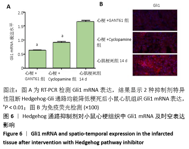
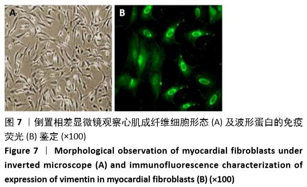
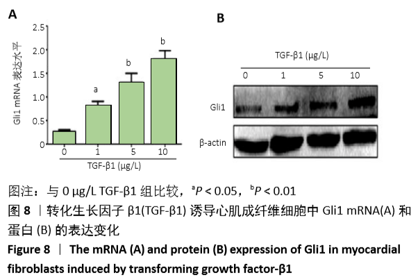
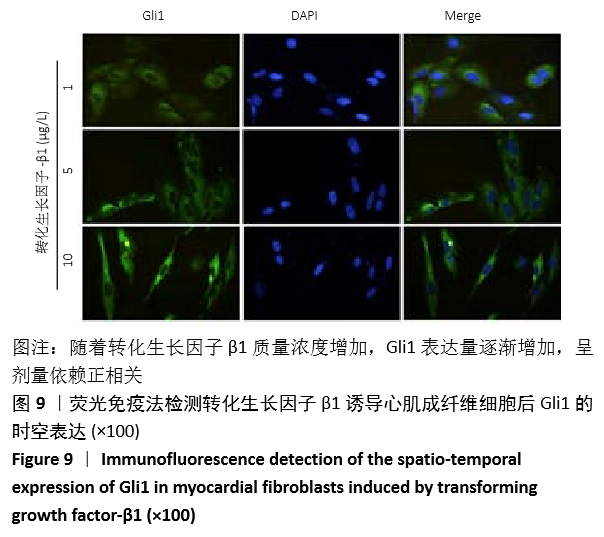
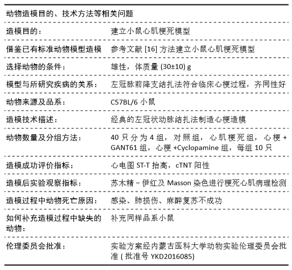
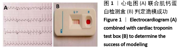
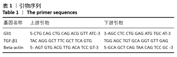
方法:①动物实验:左冠状动脉结扎法建立小鼠心肌梗死模型,苏木精-伊红和Masson染色法观察梗死后3,7,14 d心肌组织的病理改变,用RT-PCR、Western blot和免疫荧光法检测不同时段Gli1 mRNA及蛋白的时空表达,RT-PCR和免疫荧光定量检测加入特异性阻断剂后Gli1的mRNA及蛋白的时空表达变化。②细胞实验:差速贴壁法原代培养SD乳鼠心肌成纤维细胞,采用不同质量浓度(0,1,5,10 μg/L)转化生长因子β1刺激心肌成纤维细胞,RT-PCR、Western blot检测Gli1 mRNA及蛋白表达,选用10 μg/L的转化生长因子β1诱导心肌成纤维细胞24 h,免疫荧光法检测Gli1蛋白时空表达及上皮间质转分化标记物的表达。分别采用GANT61和Cyclopamine特异性阻断Hedgehog信号通路中的Gli1和Smo 24 h后,加入10 μg/L转化生长因子β1,RT-PCR检测Gli1 mRNA表达变化,荧光免疫法检测Gli1的时空表达以及上皮间质转分化标记物的表达变化。
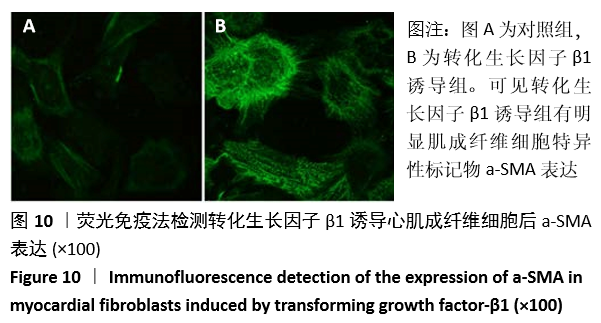
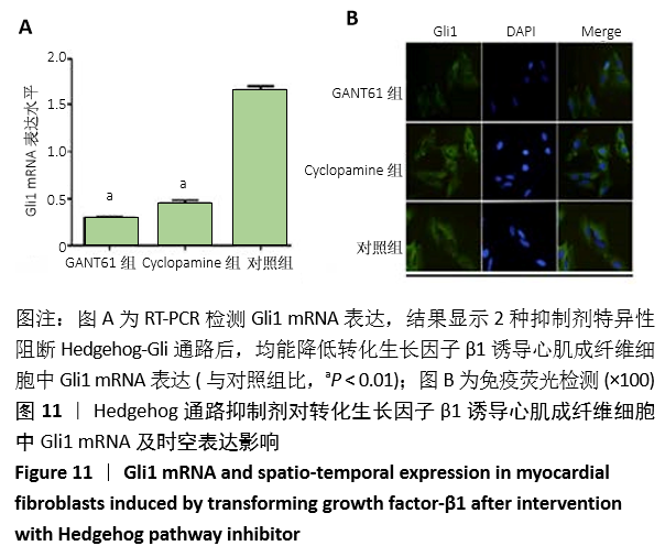
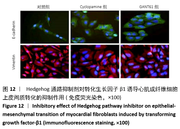
结果与结论:①动物实验结果:随着梗死后时间延长,小鼠心肌Gli1的mRNA和蛋白表达水平逐渐升高,而加入Hedgehog通路特异性阻断剂后梗死组织中Gli1的mRNA及蛋白表达均有下降。②细胞实验结果:采用转化生长因子β1刺激后,心肌成纤维细胞上皮间质转化为肌成纤维细胞(特异性标记物a-SMA阳性);随着转化生长因子β1干预剂量增加,Gli1的mRNA及蛋白表达逐渐增加。采用阻断剂特异性阻断Hedgehog信号通路中的Gli1和Smo 24 h后,Gli1的mRNA和蛋白表达均受抑制,同时伴有上皮间质转化发生的生物标记物E-Cadherin和Vimentin的表达改变,证实发生了上皮间质转化。③结合体内及体外实验,共同证实了Hedgehog-Gli信号通路参与心肌纤维化及心肌成纤维细胞转分化发生发展过程。
https://orcid.org/0000-0001-5878-9882 (聂慧娟) 

中国组织工程研究杂志出版内容重点:组织构建;骨细胞;软骨细胞;细胞培养;成纤维细胞;血管内皮细胞;骨质疏松;组织工程

关键词: 转化生长因子β1, 通路, 上皮间质转化, 心肌, 成纤维细胞, 心肌梗死, 人类间质瘤相关致癌基因同源体1

Abstract: BACKGROUND: In recent years, increasing studies have shown that abnormally activated Hedghog signaling pathway is widely involved in the injury repair of systemic multi-tissue organ diseases, but its related role in myocardial fibrosis is still unclear.
OBJECTIVE: To study the effect of blocking Hedgehog-Gli signaling pathway on epithelial-mesenchymal transition of myocardial fibroblasts induced by transforming growth factor-β1 (TGF-β1).
METHODS: (1) Animal experiment: The model of myocardial infarction in mice was established by left coronary artery ligature. The pathological changes of myocardial tissue were observed by hematoxylin-eosin staining and Masson staining at 3, 7, and 14 days after infarction. The mRNA, protein and spatio-temporal expressions of Gli1 at different time points were detected by RT-PC, western blot and immunofluorescence. The mRNA and protein expression changes of Gli1 before and after adding inhibitor were detected using RT-PCR and immunofluorescence. (2) Cell experiment: The primary cultured myocardial fibroblasts of neonatal Sprague-Dawley rats were cultured by differential adherent method. The cultured myocardial fibroblasts were cultured with 0, 1, 5, and 10 μg/L TGF-β1 intervention. The expression of Gli1 mRNA and protein was detected by RT-PCR and western blot, respectively. Myocardial fibroblasts were induced by TGF-β1 of 10 μg/L for 24 hours, and the spatio-temporal expression of Gli1 protein and expression of epithelial-mesenchymal transition markers were detected by immunofluorescence. Gli1 and Smo in Hedgehog signaling pathway were specifically blocked by GANT61 and Cyclopamine for 24 hours, respectively, and 10 μg/L TGF-β1 was added. The mRNA expression level of Gli1 was detected by RT-PCR, and the spatio-temporal expression of Gli1 and expression of epithelial-mesenchymal transition markers were detected by immunofluorescence.
RESULTS AND CONCLUSION: (1) Results of the animal experiment: As the time after infarction prolonged, the mRNA and protein expression levels of Gli1 in the mouse myocardium gradually increased, but the mRNA and protein expressions of Gli1 in the infarcted tissue decreased after addition of the Hedgehog pathway specific blocker. (2) Results of the cell experiment: After stimulation with TGF-β1, the epithelial mesenchyme of myocardial fibroblasts transformed into myofibroblasts (positive for a-SMA). As the intervention dose of TGF-β1 increased, the mRNA and protein expression of Gli1 gradually increased. After specifically blocking Gli1 and Smo in the Hedgehog signaling pathway for 24 hours, the mRNA and protein expression levels of Gli1 were inhibited, accompanied by the changes in the expression of E-Cadherin and Vimentin during epithelial-mesenchymal transition, indicating the existence of epithelial-mesenchymal transition. By the combination of in vivo and in vitro experiments, this study confirmed that the Hedgehog-Gli signal pathway is involved in the occurrence and development of myocardial fibrosis and myocardial fibroblast transdifferentiation.

Key words: transforming growth factor-β1, pathway, epithelial-mesenchymal transition, myocardium, fibroblast, myocardial infarction, human glioma associated oncogene homolog 1

中图分类号: