中国组织工程研究 ›› 2020, Vol. 24 ›› Issue (19): 3017-3022.doi: 10.3969/j.issn.2095-4344.2066

• 干细胞因子及调控因子 stem cell factors and regulatory factors • 上一篇    下一篇

碱性成纤维细胞生长因子及胰岛素样生长因子1对精原干细胞增殖凋亡影响的机制

李  宏1,2,吴绍华1,邱明星1,邓青富3,雷国林2,李青龙2   

  1. 1西南医科大学,四川省泸州市  646000;2简阳市人民医院·西南医科大学附属简阳医院泌尿外科,四川省简阳市  641400;3西南医科大学附属医院泌尿外科,四川省泸州市  646000
  • 收稿日期:2019-07-30 修回日期:2019-08-02 接受日期:2019-09-17 出版日期:2020-07-08 发布日期:2020-04-08
  • 通讯作者: 邱明星,主任医师,西南医科大学,四川省泸州市 646000
  • 作者简介:李宏,男,1980年生,四川省成都市人,副主任医师,主要从事泌尿疾病方面的研究。
  • 基金资助:
    四川省卫生和计划生育科研课题项目(17PJ160)

Mechanism underlying basic fibroblast growth factor and insulin-like growth factor-1 effects on proliferation and apoptosis of spermatogonial stem cells

Li Hong1, 2, Wu Shaohua1, Qiu Mingxing1, Deng Qingfu3, Lei Guolin2, Li Qinglong2   

  1. 1Southwest Medical University, Luzhou 646000, Sichuan Province, China; 2Department of Urology Surgery, the People’s Hospital of Jianyang City & Jianyang Hospital of Southwest Medical University, Jianyang 641400, Sichuan Province, China; 3Department of Urology Surgery, Affiliated Hospital of Southwest Medical University, Luzhou 646000, Sichuan Province, China
  • Received:2019-07-30 Revised:2019-08-02 Accepted:2019-09-17 Online:2020-07-08 Published:2020-04-08
  • Contact: Qiu Mingxing, Chief physician, Southwest Medical University, Luzhou 646000, Sichuan Province, China
  • About author:Li Hong, Associate chief physician, Southwest Medical University, Luzhou 646000, Sichuan Province, China; Department of Urology Surgery, the People’s Hospital of Jianyang City & Jianyang Hospital of Southwest Medical University, Jianyang 641400, Sichuan Province, China
  • Supported by:
    the Research Project of the Health and Family Planning Commission of Sichuan Province, No. 17PJ160

摘要:

文题释义:

碱性成纤维细胞生长因子:是细胞生长和分化的重要调节因子,具有促血管生成、细胞增殖、细胞趋化、细胞迁移等活性,在细胞分化和机体发育过程中发挥重要作用。碱性成纤维细胞生长因子通过与细胞膜表面的特异性配体结合,进而引发细胞内的一系列级联反应,从而产生各种生物学效应。

胰岛素样生长因子1:是多功能细胞增殖调控因子,主要分布在肝脏中,其与胰岛素样生长因子2、胰岛素及其受体一起构成了胰岛素样生长因子家族,其对细胞生长和代谢具有多效作用。

背景:生长因子作为体外细胞培养和体内细胞生长及增殖必需的调节因子,一直被广泛的关注。

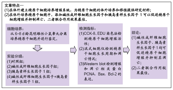
目的:探讨碱性成纤维细胞生长因子(basic fibroblast growth factor,bFGF)与胰岛素样生长因子1(insulin-like growth facter,IGF-1)联合作用对小鼠精原干细胞增殖、凋亡的影响。

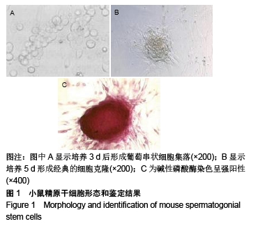
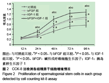
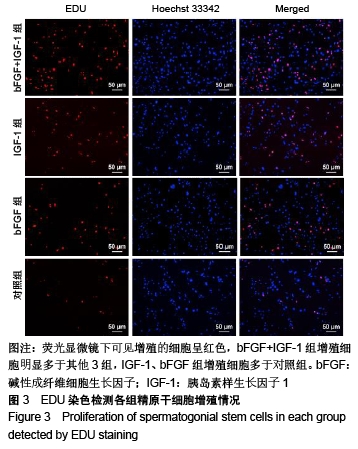
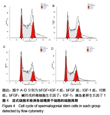
方法:从6-8 d龄昆明雄性小鼠睾丸内分离培养精原干细胞并进行鉴定。将精原干细胞接种于经丝裂霉素C处理过的胚胎成纤维细胞饲养层上,分组干预:对照组加入正常DMEM培养基进行培养;bFGF、IGF-1组分别加入含20 μg/L bFGF、20 μg/L IGF-1的DMEM培养基进行培养;bFGF+IGF-1组同时加入含20 μg/L bFGF及20 μg/L IGF-1的DMEM培养基进行培养。采用CCK-8、EDU染色法分别检测精原干细胞增殖活性,流式细胞仪检测精原干细胞生长周期和细胞凋亡情况,Western blot检测增殖和凋亡相关蛋白PCNA、Bax、Bcl-2的表达。

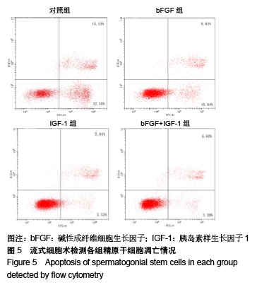
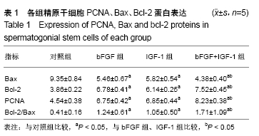
结果与结论:①与对照组比较,bFGF组、IGF-1组、bFGF+IGF-1组吸光度值显著升高,与bFGF组、IGF-1组比较,bFGF+IGF-1组吸光度值进一步升高(P < 0.05),EDU染色得到与CCK-8实验一致的结论;②bFGF+IGF-1组S+G2/M期细胞比例明显高于其他3组(P < 0.05),IGF-1组、bFGF组S+G2/M期细胞比例高于对照组(P < 0.05);③与对照组比较,bFGF组、IGF-1组、bFGF+IGF-1组凋亡细胞降低;与bFGF组、IGF-1组比较,bFGF+IGF-1组凋亡细胞进一步降低;④与对照组比较,bFGF组、IGF-1组、bFGF+IGF-1组细胞中Bax蛋白相对表达水平显著下降(P < 0.01),Bcl-2和PCNA蛋白相对表达水平均显著升高(P < 0.05)。与bFGF组、IGF-1组比较,bFGF+IGF-1组细胞中Bax蛋白相对表达水平进一步下降(P < 0.01),Bcl-2和PCNA蛋白相对表达水平进一步升高(P < 0.05);⑤结果表明,bFGF、IGF-1通过上调PCNA和Bcl-2蛋白的表达,下调Bax蛋白的表达,促进细胞增殖,抑制细胞凋亡,二者联合作用效果最佳。

ORCID: 0000-0001-5693-3713(李宏)

中国组织工程研究杂志出版内容重点:干细胞;骨髓干细胞;造血干细胞;脂肪干细胞;肿瘤干细胞;胚胎干细胞;脐带脐血干细胞;干细胞诱导;干细胞分化;组织工程

关键词: 精原干细胞, 胰岛素样生长因子1, 碱性成纤维细胞生长因子, 细胞增殖, 细胞凋亡

Abstract:

BACKGROUND: As a necessary regulatory factor of in vitro cell culture, and in vivo cell growth and proliferation, growth factors have attracted much attention.

OBJECTIVE: To investigate the effects of basic fibroblast growth factor (bFGF) combined with insulin-like growth factor-1 (IGF-1) on the proliferation and apoptosis of spermatogonial stem cells in mice.

METHODS: Spermatogonial stem cells were isolated and cultured from the testis of 6-8 days old male Kunming mice and were identified. Spermatogonial stem cells were inoculated into the feeder layer of embryonic fibroblasts treated with mitomycin C, and were then divided into four groups. Control group was cultured with normal DMEM medium; bFGF and IGF-1 groups were cultured with DMEM medium containing

20 μg/L bFGF and 20 μg/L IGF-1, respectively; bFGF+IGF-1 group was cultured with DMEM medium containing 20 μg/L bFGF and 20 μg/L IGF-1. The proliferation activity of spermatogonial stem cells was detected by cell counting kit-8 assay and EDU staining. The growth cycle and apoptosis of spermatogonial stem cells were detected by flow cytometry. The expression levels of PCNA, Bax and Bcl-2 proteins were detected by western blot assay.

RESULTS AND CONCLUSION: Compared with the control group, the absorbance values in the bFGF, IGF-1 and bFGF+IGF-1 groups were significantly increased. Compared with the bFGF and IGF-1 groups, the absorbance values in the bFGF+IGF-1 group were further increased (P < 0.05). EDU staining results showed the same conclusion as cell counting kit-8 assay results. The proportion of S+G2/M phase cells in the bFGF+IGF-1 group was significantly higher than that in the other three groups (P < 0.05). The proportion in the IGF-1 and bFGF groups was significantly higher than that in control group (P < 0.05). Compared with the control group, the number of apoptotic cells in the bFGF, IGF-1 and bFGF+IGF-1 groups was decreased. Compared with the bFGF and IGF-1 groups, the number of apoptotic cells in the bFGF+IGF-1 group was further decreased. Compared with the control group, the relative expression levels of Bax protein in the bFGF, IGF-1 and bFGF+IGF-1 groups were significantly decreased (P < 0.01), and the expression levels of Bcl-2 and PCNA proteins were significantly increased (P < 0.05). Compared with the bFGF and IGF-1 groups, the relative expression level of Bax protein in the bFGF + IGF-1 group was decreased further (P < 0.01), and the relative expression levels of Bcl-2 and PCNA proteins were increased further (P < 0.05). These results indicate that bFGF and IGF-1 can promote cell proliferation and inhibit cell apoptosis by up-regulating the expression of PCNA and Bcl-2 proteins and down-regulating the expression of Bax protein. The combination of bFGF and IGF-1 can achieve favorable effects.

Key words: spermatogonial stem cells, insulin-like growth factor 1, basic fibroblast growth factor, cell proliferation, cell apoptosis

中图分类号: