| [1] Marsboom G, Janssens S. Endothelial progenitor cells: new perspectives and applications in cardiovascular therapies.Expert Rev Cardiovasc Ther.2008;6(5): 687-701.
[2] Hattar K, Fink L, Fietzner K,et al. Cell density regulates neutrophil IL-8 synthesis: role of IL-1 receptor antagonist and soluble TNF receptors. J Immunol. 2001;166(10):6287-6293.
[3] Thomson JA, Itskovitz-Eldor J, Shapiro SS, et al. Embryonic stem cell lines derived from human blastocysts. Science.1998;282:1145-1147.
[4] Kim JH, Auerbach JM, Rodriguez JA, et al. Dopamine neurons derived fromembryonic stem cells function in an animal model of Parkinson disease. Nature. 2002; 418:50-56.
[5] Inoue M, Ino Y, Gibo J, et al. The role of MCP-1 (monocyte chemoattractant protein-1)in experimental chronic pancreatitis model induced by dibutyltin dichloride in rats. Pancreas.2002;25:e64-70.
[6] Friedenstein AJ, Gorskaja JF, Kulagina NN. Fibroblast precursors in normal and irradiated mouse hematopoietic organs. Hematol. 1976;4:267-274
[7] Pournasr B, Mohamadnejad M, Bagheri M, et al. In vitro differentiation of human bone marrow mesenchymal stem cells into hepatocyte-like cells. Archives of iranian Medicine. 2011;4(14):244-249.
[8] Tiaii H, Bharadwaj S,Liu Y, et al. Differentiation of human bone marrow mesenchymal stem cells into bladder cells: potential for urological tisstue engineering. Tissue Eng Part A.2010;16(5):1769-1779.
[9] Farrell E,Both SK. Odorfer KI, et al. In-vivo generation of bone via endochondral ossification by in-vitro chondrogenic priming of adult human and rat mesenchymal stem cells. Orthop Res.2011; 29(7): 1075-1080.
[10] Polisetti N,Chaitanya VG,Babu PP, et al. Isolation,characterization and differentiation potential of rat bone marrow stromal cells. Neurol India 2010; 58:201-208.
[11] 王丹丹,张华勇,顾志.骨髓间充质干细胞对原发性胆汁性肝硬化动物模型治疗作用研究初探[J].中华风湿病学杂志,2009,13(11): 778.
[12] 李华,乙肝患者骨髓间充质干细胞乙肝病毒感染状况的研究[M].中山大学,2007.
[13] 曾令兰,熊开钧,黄华芳,等.乙肝病毒体外感染正常人骨髓中3种细胞的动态研究[J].同济医科大学学报,1996.25(2): 128-130.
[14] 中华人民共和国科学技术部.关于善待实验动物的指导性意见.2006-09-30.
[15] 邱嘉华,陈奕金,贾林,等.牛磺胆酸钠诱导急性坏死性胰腺炎大鼠模型的理想浓度[J].中华胰腺病, 2010,10(2): 120-122.
[16] Tokalov SV,Gruener S,Schindler S, et al. A number of bone marrow mesenchymal stem but neither phenotype nor differentiation capacities changes with age of rats. Mol Cells. 2007;24(2):255-260.
[17] Hudson LD, Steinberg KP. Epidemiology o f acute lung in-jury and A RDS. Chest.1999;116:74S .
[18] Steer M. Relationship between pancreatitis and lung diseases. Respir Physiol. 2001;1 28:13 .
[19] 程石,何三光,张佳林.肺泡巨噬细胞活化在急性坏死性胰腺炎大鼠肺损伤中的作用[J]. 中华外科杂志, 2002,40(8): 609.
[20] Aho HJ, Koskensalo SM, Nevalainen TJ. Experimental pancreatitis in the rat. Sodium taurocholate-induced acute hacmorrhagic pancreatitis. Scand J Gastroenterol.1980; 15(4):411-416.
[21] 习佳飞,王锰芳,裴雪涛.成体干细胞及其在再生医学中的应用[J].生命科学,2006,18:328-332.
[22] Tamir APT, Stetler K, Stetler K,et al.Stem eell faetor inhibits erythroid differentiations by modulating the activity of G1 cyclin dependent kinase complexes: a role for P27 in erythroid differentiation coupled G1 arrest .CellGrowth.2000:11:269-277.
[23] Quirici N, Soligo D , Bossolaseo P , et al. Isolation of bone marrow mesenchymal stem cells by anti-nerve growth factor receptor antibodies. Exp Hematol.2002: 30(7):78-791.
[24] Rojas M, Xu J. Bone marrow-derived mesenehymal stem cells in repair of the injured lung.Am J Respir Cell Mol Biol.2005;33:145-152.
[25] 江学良,李兆申,崔慧斐.骨髓间充质干细胞在胰腺生理更新和病理再生中的作用[J].世界华人消化杂志,2006: 14(4): 398-404.
[26] Cui HF, Bai ZL. Protective effects of transplanted and mobilized bone marrow stem cells on mice with severe acute panereatitis. World J Gastroenterol.2003; 9(10): 2274-2277.
[27] 王欣燕, 吴晓梅,康小文,等.骨髓间质干细胞移植对博莱霉素诱导大鼠肺纤维化的治疗作用[J].中国呼吸与危重监护杂志,2009,1(8):57-62.
[28] 李奇智,吴新民,郭亚民.相关细胞因子在重症急性胰腺炎相关肺损伤中的作用[J].中国普外基础与临床杂志,2011, 3(18):258-260.
[29] 姜战武,李厚祥,邵苏吉.血清PLA2对急性胰腺炎的诊断和预后判断价值[J].南通医学院学报,1996,16(3): 326-327.
[30] 付林,程书榜,高海斌,等. IL-10区域动脉灌注减轻急性胰腺炎大鼠肺损伤的实验研究[J].中国普通外科杂志, 2006, 15(8): 619-621.
[31] 莫坚,刘欣,何海燕,等.倍他米松对大鼠重症急性胰腺炎肺损伤NF-JB、ICAM-1及IL-6的影响[J].湖北民族学院学报:医学版, 2005,22(4):14-16.
[32] Seamas C, Strieter RM , Reid PT, et al. The association between mortality rates and decreased concent rations of interleukin- 10 and interleukin- 1 receptor antagonist in the lung fluids of patient s with the adult respiratory distress syndrome. Ann Intern Med.1996;125(3):191.
[33] Leme AS, Lichtenstein A, Arantes Costa FM, et al. Acute lung injury in experimental pancreatitis in rats: pulmonary protective effects of crotapotin and N-acetylcysteine.Shock.2002;18(5):428-433.
[34] 陆海华,史忠,刘波,等.急性胰腺炎相关性肺损伤早期血清TNF-α、IL-1、IL-6的变化及其临床意义[J].重庆医学,2007, 36(18): 1818-1819.
[35] Altavilla D, Famulari C, Passaniti M, et al. Lipid peroxidation inhibition reduces NF-JB activation and attenuates cerulein-induced pancreatitis. Free Radic Res.2003; 37(4):425-435.
[36] 周粼,梁道明,陈嘉勇,等.依达拉奉对大鼠重症急性胰腺炎肺损伤的影响[J].中国普外基础与临床杂志, 2010,17(2): 125-154. |
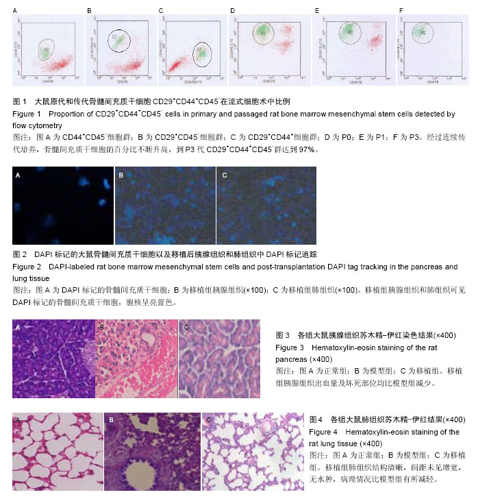
.jpg)
.jpg)