| [1] Li C, Wei G, Gu Q,et al.Donor Age and Cell Passage Affect Osteogenic Ability of Rat Bone Marrow Mesenchymal Stem Cells. Cell Biochem Biophys.2015 Jan 30. [Epub ahead of print]
[2] 李嘉雯,王晓云,简白羽,等.间充质干细胞的临床应用与问题研究[J].医学综述, 2015, 21(4): 613-615.
[3] Roberts SJ, van Gastel N, Carmeliet G, et al. Uncovering the periosteum for skeletal regeneration: the stem cell that lies beneath. Bone.2015;70:10-18.
[4] Tang Y, Wang B.Gene- and stem cell-based therapeutics for cartilage regeneration and repair. Stem cell research & therapy.2015;6(1): 78.
[5] Jansen EJ, Emans PJ, Guldemond NA, et al. Human periosteum-derived cells from elderly patients as a source for cartilage tissue engineering? . J Tissue Eng Regen Med. 2008;2(6):331-339.
[6] Samee M, Kasugai S, Kondo H, et al.Bone morphogenetic protein-2 (BMP-2) and vascular endothelial growth factor (VEGF) transfection to human periosteal cells enhances osteoblast differentiation and bone formation.J Pharmacol Sci. 2008;108(1):18-31.
[7] Ichijima T, Matsuzaka K, Tonogi M, et al. Osteogenic differences in cultured rat periosteal cells under hypoxic and normal conditions.Exp Ther Med. 2012; 3(2):165-170.
[8] Castro-Silva II, Zambuzzi WF, de Oliveira Castro L, et al.Periosteal-derived cells for bone bioengineering: a promising candidate. Clin Oral Implants Res. 2012; 23(10):1238-1242.
[9] Caterson EJ, Nesti LJ, Danielson KG, et al.Human marrow-derived mesenchymal progenitor cells: isolation, culture expansion, and analysis of differentiation.Mol biotechnol.2002; 20(3): 245-256.
[10] Hah YS, Joo HH, Kang YH, et al.Cultured human periosteal-derived cells have inducible adipogenic activity and can also differentiate into osteoblasts in a perioxisome proliferator-activated receptor-mediated fashion.Int J Med Sci. 2014;11(11):1116-1128.
[11] Sheng G. The developmental basis of mesenchymal stem/stromal cells (MSCs). BMC Developmental Biology.2015;15(1): 44.
[12] Yu T, Volponi AA, Babb R, An Z, et al.Stem Cells in Tooth Development, Growth, Repair, and Regeneration. Curr Top Dev Biol.2015;115:187-212.
[13] Chen H, Niu JW, Ning HM, et al.Treatment of Psoriasis with Mesenchymal Stem Cells. Am J Med. 2016;129(3): e13-4
[14] van Gastel N,Torrekens S,Roberts SJ et al.engineering vascularized bone: osteogenic and proangiogenic potential ofmurine periosteal cells.Stem Cells.2012; 30(11):2460-71
[15] 杨宇明,袁峰,陆海涛,等. 骨膜细胞与髓核细胞共培养向成骨方向的分化[J]. 中国组织工程研究,2015,19(37): 5916-5922.
[16] Bei K, Du Z, Xiong Y, et al. BMP7 can promote osteogenic differentiation of human periosteal cells in vitro. Molecular biology reports.2012; 39(9): 8845-8851.
[17] 贝抗胜,孙庆文,熊英辉,等.成骨因子 BMP7 在骨膜细胞体外培养中的作用[J].中华显微外科杂志,2010,33(5). 384-387.
[18] Arnsdorf EJ, Jones LM, Carter DR,et al. The periosteum as a cellular source for functional tissue engineering. Tissue Eng Part A 2009;9: 2637-2642
[19] [19] Hutmacher DW, Sittinger M. Periosteal cells in bone tissue engineering. Tissue Eng.2003;9(15): S45-S64.
[20] Yu Y Y, Lieu S, Lu C,et al.Bone morphogenetic protein 2 stimulates endochondral ossification by regulating periosteal cell fate during bone repair. Bone.2010; 47(1): 65-73.
[21] Bolander ME. Regulation of fracture repair by growth factors. Proceedings of the Society for Experimental Biology and Medicine Society for Experimental Biology and Medicine (New York, NY).1992;200(2):165-170.
[22] Deren JA, Kaplan FS, Brighton CT. Alkaline phosphatase production by periosteal cells at various oxygen tensions in vitro. Clin Orthop Relat Res. 1990; (252):307-312.
[23] 贝抗胜,吴礼杨,孙庆文,等.人骨膜细胞生物学特性的实验研究[J]. 中华创伤骨科杂志, 2011, 13(12):1170-1174.
[24] Choi YS, Noh SE, Lim SM, et al. Multipotency and growth characteristic of periosteum-derived progenitor cells for chondrogenic, osteogenic, and adipogenic differentiation. Biotechnology letters.2008;30(4): 593-601.
[25] Ball M D, Bonzani I C, Bovis M J, et al. Human periosteum is a source of cells for orthopaedic tissue engineering: a pilot study. Clin Orthop Relat Res. 2011; 469(11):3085-3093.
[26] Yoshimura H, Muneta T, Nimura A, et al. Comparison of rat mesenchymal stem cells derived from bone marrow, synovium, periosteum, adipose tissue, and muscle. Cell Tissue Res. 2007;327(3):449-462.
[27] Dominici M, Le Blanc K, Mueller I, et al.Minimal criteria for defining multipotent mesenchymal stromal cells. Cytotherapy.2006;8(4):315-317.
[28] Kobolak J, Dinnyes A, Memic A, et al. Mesenchymal stem cells: Identification, phenotypic characterization, biological properties and potential for regenerative medicine through biomaterial micro-engineering of their niche. Methods. 2016;99:62-68.
[29] De Bari C, Dell'Accio F, Vanlauwe J, et al. Mesenchymal multipotency of adult human periosteal cells demonstrated by single-cell lineage analysis. Arthritis Rheum. 2006 ;54(4):1209-1221.
[30] Yoshimura H, Muneta T, Nimura A, et al. Comparison of rat mesenchymal stem cells derived from bone marrow, synovium, periosteum, adipose tissue, and muscle. Cell Tissue Res.2007; 327(3): 449-462.
[31] Almalki SG, Agrawal DK. Key transcription factors in the differentiation of mesenchymal stem cells. Differentiation. 2016 Mar 21. pii: S0301-4681(15) 30098-0.
[32] Friedenstein AJ, Petrakova KV, Kurolesova AI, et al.Heterotopic of bone marrow. Analysis of precursor cells for osteogenic and hematopoietic tissues. Transplantation.1968;6(2):230-247. |
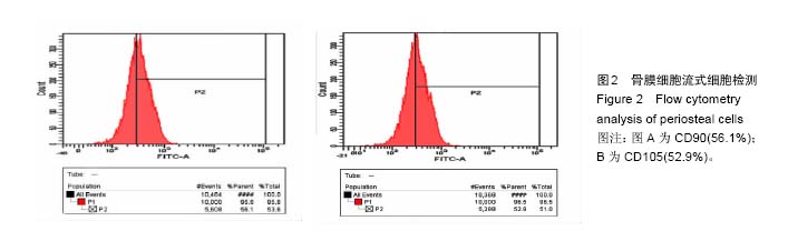
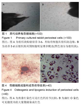
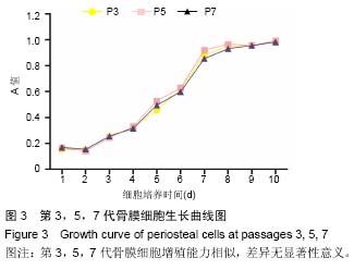
.jpg)
.jpg)