中国组织工程研究 ›› 2015, Vol. 19 ›› Issue (40): 6460-6464.doi: 10.3969/j.issn.2095-4344.2015.40.012

• 器官移植动物模型 organ transplantation and animal model • 上一篇    下一篇

高压氧治疗促进脑梗死模型大鼠神经再生微环境及神经功能的恢复

孙晶晶1,宋乃光2,张耀龙2,高淑焕1,孙彩悦1,薛 建1,贺永贵3,习瑾琨3,张国彬3   

  1. 唐山市协和医院,1神经内一科,2急诊科,河北省唐山市  063000;3河北联合大学培养办公室,河北省唐山市  063000
  • 出版日期:2015-09-30 发布日期:2015-09-30
  • 通讯作者: 张国彬,硕士,河北联合大学培养办公室,河北省唐山市 063000
  • 作者简介:孙晶晶,女,1979出生,天津市人,汉族, 2004年河北医科大学毕业,硕士,主治医生

Hyperbaric oxygen therapy improves nerve regeneration microenvironment and promotes rat nerve function recovery after cerebral infarction 

Sun Jing-jing1, Song Nai-guang2, Zhang Yao-long2, Gao Shu-huan1, Sun Cai-yue1, Xue Jian1He Yong-gui3, Xi Jin-kun3, Zhang Guo-bin3   

  1. 1First Department of Neurology, 2Department of Emergency, Tangshan Union Medical College Hospital, Tangshan 063000, Hebei Province, China; 3Training Office, Hebei United University, Tangshan 063000, Hebei Province, China
  • Online:2015-09-30 Published:2015-09-30
  • Contact: Zhang Guo-bin, Master, Training Office, Hebei United University, Tangshan 063000, Hebei Province, China
  • About author:Sun Jing-jing, Master, Attending physician, First Department of Neurology, Tangshan Union Medical College Hospital, Tangshan 063000, Hebei Province, China

摘要:

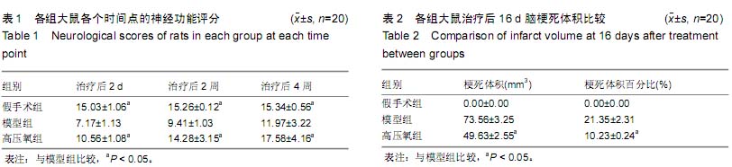
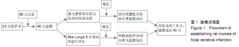
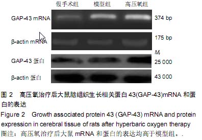
背景:大量临床研究证实,脊髓损伤区域的微环境通过高压氧可获得显著改善,但高压氧对脑损伤微环境有何影响呢?
目的:观察高压氧治疗对大鼠脑梗死区神经再生微环境及神经功能恢复的影响。
方法:构建大脑中动脉阻断制作局灶性脑梗死模型大鼠,并进行高压氧治疗,设假手术组和模型组作对照,假手术组不结扎颈内动脉不干预,模型组造模后模拟除压力和氧浓度以外的其他高压氧治疗过程及环境条件。
结果与结论:与模型组相比,治疗后第16天高压氧组的大鼠肢体功能评分、术后14 d 脑梗死区组织生长相关蛋白43表达均升高(P < 0.05),术后24 h梗死灶体积均降低(P < 0.05)。结果证实,高压氧治疗促进脑梗死模型大鼠神经再生微环境及神经功能的恢复。

 中国组织工程研究杂志出版内容重点:肾移植肝移植移植;心脏移植;组织移植;皮肤移植;皮瓣移植;血管移植;器官移植组织工程

关键词: 实验动物, 脑及脊髓损伤动物模型, 脑梗死, 缺血性, 大鼠, 高压氧, 神经再生, 运动功能, 生长相关蛋白43, 微环境, 恢复, 细胞

Abstract:

BACKGROUND: Numerous clinical studies have confirmed that the microenvironment at a spinal cord injury site can be obviously improved through hyperbaric oxygen therapy; however, what effect does hyperbaric oxygen  have on the microenvironment of the injured brain?

OBJECTIVE: To investigate the effect of hyperbaric oxygen therapy on nerve regeneration microenvironment and the recovery of rat nerve function after focal cerebral infarction.

METHODS: Rat models of focal cerebral infarction were established by occlusion of the middle cerebral artery and subjected to hyperbaric oxygen therapy. Sham group and model group were established as comparison. In the sham group, rat models of focal cerebral infarction were established but did not receive any treatment. Rats in the model group were placed in a hyperbaric oxygen therapy chamber but the pressure and oxygen concentration were not administered.  

RESULTS AND CONCLUSION: Compared with the model group, the score of rat limb function at 16 days after treatment and the expression of growth associated protein 43 in the rat cerebral infarcted area at postoperative 14 days were significantly increased , but infarct volume at postoperative 24 hours was all significantly decreased in the hyperbaric oxygen therapy group (all P < 0.05). These results confirmed that hyperbaric oxygen therapy can improve nerve regeneration microenvironment and promote the recovery of rat nerve function after focal cerebral infarction.

 中国组织工程研究杂志出版内容重点:肾移植肝移植移植;心脏移植;组织移植;皮肤移植;皮瓣移植;血管移植;器官移植组织工程

中图分类号: