中国组织工程研究 ›› 2021, Vol. 25 ›› Issue (8): 1172-1176.doi: 10.3969/j.issn.2095-4344.3036

• 脊柱组织构建 spinal tissue construction • 上一篇    下一篇

不同静水压下人椎间盘髓核细胞的形态和活性

刘志超1,2,张  帆3,孙  旗3,康晓乐3,袁巧妹3,柳根哲4,陈  江3,5   

  1. 1中国中医科学院广安门医院南区骨一科,北京市   102600;2北京中医药大学,北京市   100029;3北京中医药大学东直门医院骨伤科一区,北京市   100700;4首都医科大学附属北京中医医院骨伤科,北京市   100010;5湖南中医药大学,湖南省长沙市   410208
  • 收稿日期:2020-06-06 修回日期:2020-06-12 接受日期:2020-07-11 出版日期:2021-03-18 发布日期:2020-12-10
  • 通讯作者: 陈江,博士,副主任医师,北京中医药大学东直门医院,北京市 100700;湖南中医药大学,湖南省长沙市 410208
  • 作者简介:刘志超,男,1994年生,河南省开封市人,2019年北京中医药大学毕业,硕士,主要从事中医骨伤研究。
  • 基金资助:
    国家自然科学基金项目(81603638);中国博士后科学基金面上项目(2019M662791);中央高校基本科研业务费专项资金资助(2018-JYB-XJQ010);北京中医药大学2017年度基本科研业务费项目(2017-JYB-JS-085);北京中医药大学2019年度基本科研业务费项目(2019-JYB-JS-042);北京中医大学东直门医院青苗人才项目(DZMYS-201702)

Morphology and activity of human nucleus pulposus cells under different hydrostatic pressures

Liu Zhichao1, 2, Zhang Fan3, Sun Qi3, Kang Xiaole3, Yuan Qiaomei3, Liu Genzhe4, Chen Jiang3, 5   

  1. 1Department of Orthopedics, Southern Branch of Guanganmen Hospital, China Academy of Chinese Medical Sciences, Beijing 102600, China; 2Beijing University of Chinese Medicine, Beijing 100029, China; 3Department of Orthopedics, Dongzhimen Hospital, Beijing University of Chinese Medicine, Beijing 100700, China; 4Department of Orthopedics, Beijing Traditional Chinese Medicine Hospital, Capital Medical University, Beijing 100010, China; 5Hunan University of Chinese Medicine, Hunan Province, China
  • Received:2020-06-06 Revised:2020-06-12 Accepted:2020-07-11 Online:2021-03-18 Published:2020-12-10
  • Contact: Chen Jiang, MD, Associate chief physician, Department of Orthopedics, Dongzhimen Hospital, Beijing University of Chinese Medicine, Beijing 100700, China; Hunan University of Chinese Medicine, Changsha 410208, Hunan Province, China
  • About author:Liu Zhichao, Master, Department of Orthopedics, Southern Branch of Guanganmen Hospital, China Academy of Chinese Medical Sciences, Beijing 102600, China; Beijing University of Chinese Medicine, Beijing 100029, China
  • Supported by:
    the National Natural Science Foundation of China, No. 81603638; General Program of China Postdoctoral Science Foundation, No. 2019M662791; Fundamental Research Funds for the Central Universities, No. 2018-JYB-XJQ010; 2017 Basic Scientific Research Business Expenses Project of Beijing University of Chinese Medicine, No. 2017-JYB-JS-085; 2019 Basic Scientific Research Business Expenses Project of Beijing University of Chinese Medicine, No. 2019-JYB-JS-042; Beijing Youth Talent Project of Dongzhimen Hospital of Beijing University of Chinese Medicine, No. DZMYS-201702 

摘要:

文题释义:
髓核:髓核在抵抗脊柱和椎间盘的负荷方面起着关键作用,其作用是产生一个静水压力,以均匀地分散圆盘内的负载。
静水压:是在人体内某部位聚集的液体受到应力刺激时对周围组织形成的压强。而椎间盘在人体运动时受到压应力在髓核中形成静水压,然后扩散至纤维环,由压应力转变成张应力。人平卧休息位时静水压最低约 0.3 MPa,坐位时接近 1 MPa, 弯腰搬重物可达 3 MPa。

背景:压应力能够引起细胞形态及活性的改变,但静水压下髓核细胞的形态和活性是否改变仍需进一步研究。
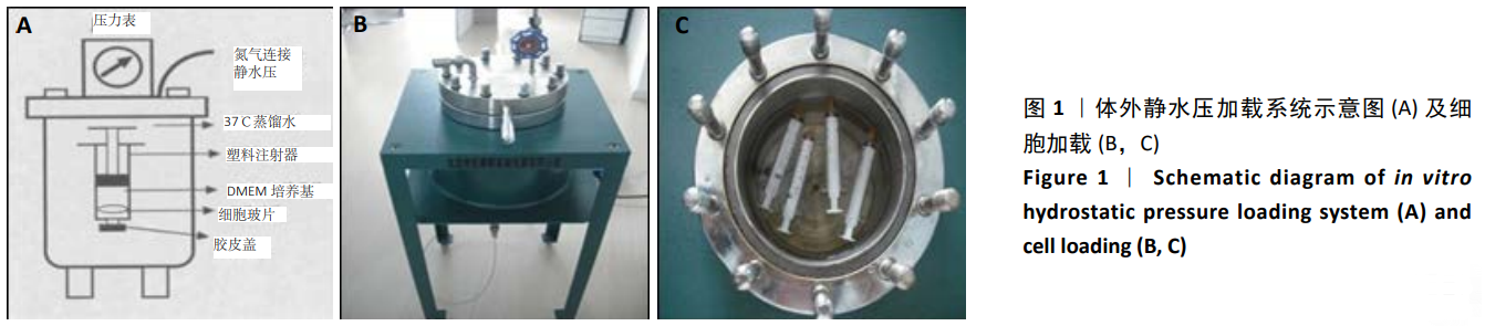
目的:研究体外静水压加载系统内人椎间盘髓核细胞形态和活性的变化。
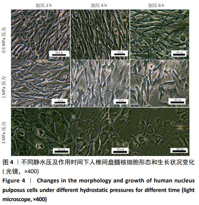
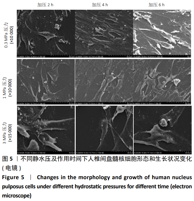
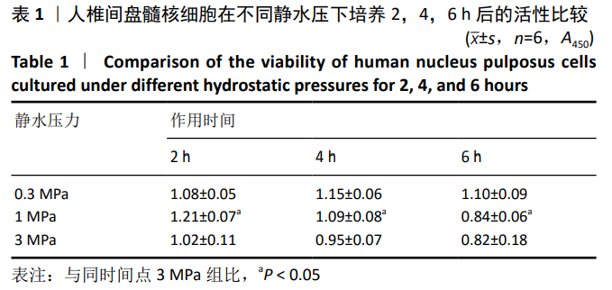
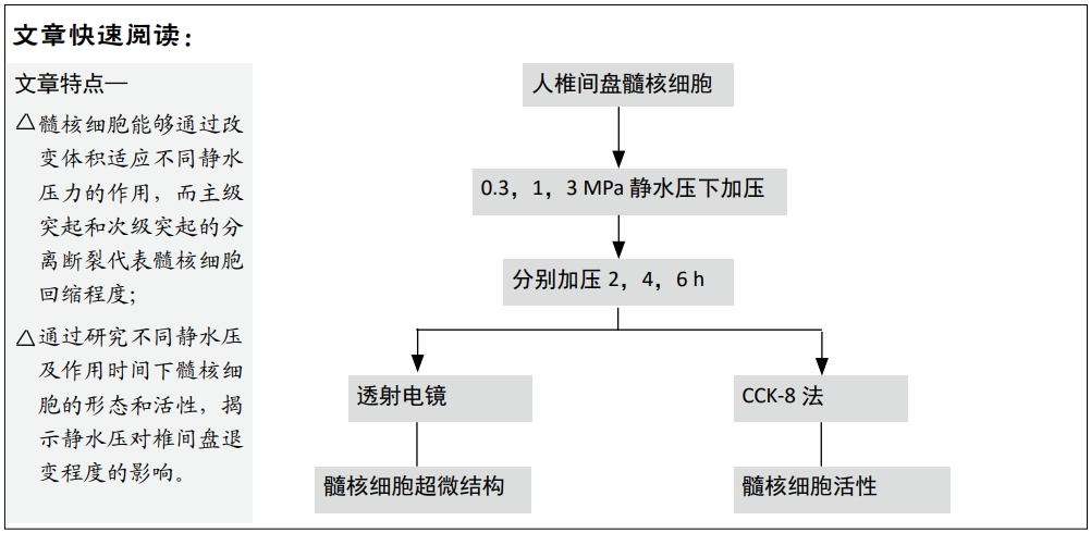
方法:将人椎间盘髓核细胞分离培养并传3代后,在静水压加载系统0.3,1,3 MPa的静水压力环境下加压2,4,6 h后,使用倒置相差显微镜观察人椎间盘髓核细胞加压前后的形态变化及生长状况;采用透射电镜观察各组人椎间盘髓核细胞超微结构的变化及差异;使用CCK-8法检测不同静水压下人椎间盘髓核细胞的增殖活性的变化。
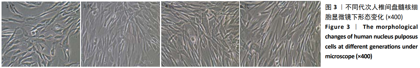
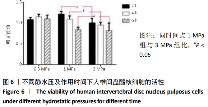
结果与结论:①细胞培养及传代:第1,2,3代人椎间盘髓核细胞的生长曲线呈S形,3-7 d时呈直线增殖,此阶段细胞生长最快、活力最高;第5,6代人椎间盘髓核细胞的突起呈长梭形,成长较慢,出现退化;②细胞形态:在0.3,1,3 MPa的静水压下人椎间盘髓核细胞均缩小,0.3,1 MPa下细胞细微变小,形态基本完整;3 MPa静水压下细胞缩小最明显,且细胞形态不完整,说明人椎间盘髓核细胞在0.3,1,3 MPa的静水压下加压2,4,6 h后,细胞形态在3 MPa静水压下改变最明显,而相同静水压下作用不同时间后细胞形态无明显改变;③细胞活性:在0.3 MPa静水压下,人椎间盘髓核细胞的增殖率随着时间的增加先升后降,4 h时细胞增殖率最高;在1,3 MPa静水压下随着时间的增加人椎间盘髓核细胞增殖率逐渐下降,且1 MPa静水压下细胞增殖率明显高于同一作用时间3 MPa静水压下(P < 0.05),说明适当的静水压力刺激有助于促进人椎间盘髓核细胞增殖,而长期不当过高的静水压刺激能使人椎间盘髓核细胞增殖率下降,导致椎间盘退变的发生。

https://orcid.org/0000-0002-6713-9061 (刘志超)

中国组织工程研究杂志出版内容重点:组织构建;骨细胞;软骨细胞;细胞培养;成纤维细胞;血管内皮细胞;骨质疏松;组织工程

关键词: 静水压, 椎间盘, 髓核细胞, 形态, 细胞活性, 生长动力学

Abstract:

BACKGROUND: Compressive stress can change the morphology and activity of cells, but whether the morphology and activity of nucleus pulposus cells change under hydrostatic pressure still needs further study.

OBJECTIVE: To study the morphology and activity of human nucleus pulposus cells in vitro. 
METHODS: The human nucleus pulposus cells were separated, cultured and passed on for three generations, and pressurized for 2, 4 and 6 hours under the hydrostatic pressure of 0.3, 1, and 3 MPa. Then, the morphological changes and growth of the cells before and after pressurization were observed by inverted phase contrast microscope. Transmission electron microscope was used to observe the ultrastructural changes and differences of the cells. Cell counting kit-8 was used to detect the proliferation activity, morphology and activity of the cells under different hydrostatic pressures. 
RESULTS AND CONCLUSION: (1) Cell culture and passage: The growth curves of the first, second and third generations of human nucleus pulposus cells were S-shaped, and the cells proliferated fastest in a straight line from 3 to 7 days. The protuberances of the 5th and 6th generation cells were long shuttle shaped, grew slowly and degenerated. (2) Cell morphology: the human nucleus pulposus cells were shrunk under hydrostatic pressures of 0.3, 1, 3 MPa. At 0.3 and 1 MPa, the cells became slightly smaller and the morphology was basically complete; at 3 MPa, the cells were most obviously shrunk and the morphology was incomplete. The results showed that when the human nucleus pulposus cells were pressurized for 2, 4 and 6 hours under 0.3, 1 and 3 MPa hydrostatic pressures, the change of cell morphology was the most obvious under 3 MPa hydrostatic pressure, but there was no obvious change under the same hydrostatic pressure for different time. (3) Cell viability: Under 0.3 MPa hydrostatic pressure, the proliferation rate of human nucleus pulposus cells first increased and then decreased with the increase of time, and the cell proliferation rate reached the peak at 4 hours. Under 1 and 3MPa hydrostatic pressures, the proliferation rate of the cells gradually decreased with the increase of time, and the cell proliferation rate under 1 MPa hydrostatic pressure was significantly higher than that under 3 MPa hydrostatic pressure at the same action time (P < 0.05). These findings indicate that proper hydrostatic pressure stimulation helps to promote the proliferation of human nucleus pulposus cells, and long-term improperly high hydrostatic pressure stimulation can reduce the proliferation rate of human nucleus pulposus cells, leading to the occurrence of intervertebral disc degeneration.

Key words: hydrostatic pressure, intervertebral disc, nucleus pulposus cells, morphology, cell viability, growth kinetics

中图分类号: