中国组织工程研究 ›› 2015, Vol. 19 ›› Issue (30): 4769-4773.doi: 10.3969/j.issn.2095-4344.2015.30.003

• 纳米生物材料 nanobiomaterials • 上一篇    下一篇

纳米羟基磷灰石/聚酰胺材料的体内成骨能力

徐显春1,王 治2,侯铁奇3   

  1. 1宜宾市第一人民医院骨一科,四川省宜宾市 644000; 2泸州医学院附属医院骨与关节外科,四川省泸州市 646000; 3暨南大学医学院,广东省广州市 510632
  • 出版日期:2015-07-16 发布日期:2015-07-16
  • 作者简介:徐显春,男,1978年生,四川省安岳县人,硕士,主要从事骨与关节损伤研究。

In vivo osteogenic capability of nano-hydroxyapatite/polyamide composite material

Xu Xian-chun1, Wang Zhi2, Hou Tie-qi3   

  1. 1First Department of Orthopedics, First People’s Hospital of Yibin, Yibin 644000, Sichuan Province, China; 2Department of Bone and Joint Surgery, Affiliated Hospital of Sichuan Medical University, Luzhou 646000, Sichuan Province, China; Medical School of Jinan University, Guangzhou 510632, Guangdong Province, China
  • Online:2015-07-16 Published:2015-07-16
  • About author:Xu Xian-chun, Master, First Department of Orthopedics, First People’s Hospital of Yibin, Yibin 644000, Sichuan Province, China

摘要:

背景:作为骨修复重建材料,纳米羟基磷灰石具有良好生物相容性及骨传导性,但临床上中单一使用纳米羟基磷灰石尚存在许多不足。

目的:观察纳米羟基磷灰石/聚酰胺材料的体内成骨能力。

方法:取24只新西兰大白兔,进行纳米羟基磷灰石/聚酰胺人工肱骨头置换,分别于置换后3,6,12,24周,进行X射线观察、组织学观察。

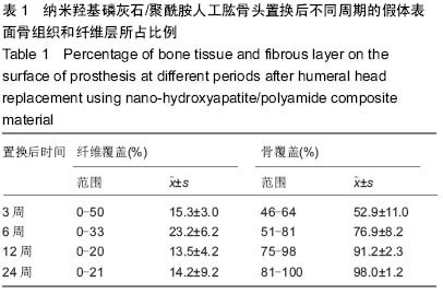
结果与结论:①X射线观察:不同时间点材料上端皮质均未出现骨皮质变薄、异位骨化等发生,纳米羟基磷灰石/聚酰胺材料无碎裂迹象,材料四周皮质均可见模糊界面,密度随着时间的增加而增加。②组织学观察:置换后3周,可见大量细胞,包括间充质细胞、单核巨噬细胞等;置换后6周,仍可见界膜内的大量纤维组织、成纤维细胞、单核巨噬细胞,而软骨细胞和成骨细胞分布较少;置换后12周,大范围原始骨小梁开始形成且多呈扁平状,排列整齐有序;置换后24周,组织界膜被骨细胞充斥,骨小梁表面的细胞较规则,骨组织原始细胞开始转变为板层状骨。说明纳米羟基磷灰石/聚酰胺材料具有良好的成骨能力。

;骨生物材料; 口腔生物材料; 纳米材料; 缓释材料; 材料相容性;组织工程

关键词: 生物材料, 骨生物材料, 纳米羟基磷灰石, 聚酰胺, 成骨能力, 广东省自然科学基金

Abstract:

BACKGROUND: As a bone reconstruction material, nano-hydroxyapatite has good biocompatibility and osteoconduction, but the clinical use of nano-hydroxyapatite alone still has many deficiencies.

OBJECTIVE: To explore the in vivo osteogenic capability of nano-hydroxyapatite/ polyamide composites.

METHODS: Twenty-four New Zealand white rabbits were subjected to humeral head replacement using nano-hydroxyapatite/polyamide composite material. X-ray observation and histological observation were done at 3, 6, 12, 24 weeks after replacement.

RESULTS AND CONCLUSION: (1) X-ray observation: No thinned cortical bone and ectopic ossification occurred on the upper end of the composite material at different time, and the nano-hydroxyapatite/polyamide material had no signs of fragmentation. The cortical bone around the composite material was fuzzy, and the bone mineral density was increased with time. (2) Histological observation: At 3 weeks after replacement, a large number of cells could be visible, including mesenchymal stem cells and mononuclear macrophages. At 6 weeks after replacement, a large amount of fibrous tissues, fibroblasts and mononuclear macrophages still existed in the boundary membrane, but chondrocytes and osteoblasts distributed less. At 12 weeks after replacement, a wide range of original trabecular bone began to form and were mostly flat that arranged regularly. At 24 weeks after replacement, the boundary membrane was full of bone cells, but the cells on the surface of trabecular bone were relatively regular and primitive cells in the bone tissue began to transform into the lamellar bone. These findings indicate that the nano-hydroxyapatite/polyamide material has good osteogenic capability

中国组织工程研究杂志出版内容重点:生物材料;骨生物材料; 口腔生物材料; 纳米材料; 缓释材料; 材料相容性;组织工程

Key words: Biocompatible Materials, Durapatite, Nylons

中图分类号: