中国组织工程研究 ›› 2015, Vol. 19 ›› Issue (28): 4435-4438.doi: 10.3969/j.issn.2095-4344.2015.28.002

• 骨髓干细胞 bone marrow stem cells • 上一篇    下一篇

骨髓间充质干细胞复合可注射型geneX及转化生长因子β2的成骨诱导效应

蓝 天,李 彪,龚跃昆,李多玉,毕 鑫,杨 毅   

  1. 昆明医科大学,云南省昆明市  650000
  • 出版日期:2015-07-02 发布日期:2015-07-02
  • 通讯作者: 李彪,博士,副主任医师,硕士生导师,昆明医科大学,云南省昆明市 650000
  • 作者简介:蓝天,男,1988年生,云南省昆明市人,在读硕士,主要从事骨关节外科研究。

Osteogenic induction of injectable geneX/bone marrow mesenchymal stem cells/ transforming growth factor beta 2 composite

Lan Tian, Li Biao, Gong Yue-kun, Li Duo-yu, Bi Xin, Yang Yi   

  1. Kunming Medical University, Kunming 650000, Yunnan Province, China
  • Online:2015-07-02 Published:2015-07-02
  • Contact: Li Biao, M.D., Associate chief physician, Master’s supervisor, Kunming Medical University, Kunming 650000, Yunnan Province, China
  • About author:Lan Tian, Studying for master’s degree, Kunming Medical University, Kunming 650000, Yunnan Province, China

摘要:

 背景:单一支架材料在不同程度不同范围内存在一些缺点,因此近年来研究者选择复合支架材料,将两种或多种具有一定互补特性的生物材料按一定方式与比例复合,构造出新型复合材料。

目的:探讨可注射型geneX人工骨复合骨髓间充质干细胞/转化生长因子β2的组织相容性及体外成骨诱导分化效果。
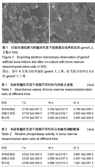
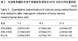
方法:将可注射型geneX人工骨与兔骨髓间充质干细胞复合培养5 d,电镜观察人工骨的组织相容性。取第3代成骨诱导培养的兔骨髓间充质干细胞分3组培养,单纯细胞组仅加入成骨诱导分化培养基,细胞支架组加入geneX人工骨与成骨诱导分化培养基,联合组加入geneX人工骨、转化生长因子β2与成骨诱导分化培养基,培养7,14,21 d进行细胞形态学观察、碱性磷酸酶活性检测、MTT检测、甲基百里香酚蓝检测及茜素红染色观察。
结果与结论:骨髓间充质干细胞在可注射型geneX人工骨表面贴壁生长,呈成纤维细胞形态,细胞基质分泌旺盛。联合组细胞增殖、成骨诱导活性强于单纯细胞组、细胞支架组(P < 0.05),细胞碱性磷酸酶活性高于单纯细胞组、细胞支架组。表明可注射型geneX人工骨复合骨髓间充质干细胞/转化生长因子β2具有良好组织相容性及促成骨分化能力。

 中国组织工程研究杂志出版内容重点:干细胞;骨髓干细胞;造血干细胞;脂肪干细胞;肿瘤干细胞;胚胎干细胞;脐带脐血干细胞;干细胞诱导;干细胞分化;组织工程

关键词: 干细胞, 骨髓干细胞, 可注射型geneX人工骨, 骨髓间充质干细胞, 转化生长因子β2, 成骨诱导, 组织相容性

Abstract:

 BACKGROUND: Single scaffold materials have some shortcomings to some extent or in different ranges. Therefore, new composite materials are developed in recent year, which are compounded by two or more materials with complementary characteristics in a certain manner or ratio.

OBJECTIVE: To investigate the histocompatibility and osteogenic induction of geneX artificial bone combined with bone marrow mesenchymal stem cells and transforming growth factor beta 2 (TGF-β2).
METHODS: Injectable geneX was co-cultured with bone marrow mesenchymal stem cells for 5 days, and under the electron microscope, the histocompatibility of the artificial bone was observed. Passage 3 rabbit bone marrow mesenchymal stem cells were divided into three groups: simple cell group cultured with osteogenic medium, cell scaffold group cultured with geneX+osteogenic medium, combined group cultured with geneX+TGF-β2+ osteogenic medium. After 7, 14, 21 days, cell morphology, alkaline phosphatase activity detection, MTT detection, methyl thymol blue detection and alizarin red staining were performed.
RESULTS AND CONCLUSION: After osteogenic induction, bone marrow mesenchymal stem cells were fibroblast-like cells and adhered to the surface of geneX bone with strong secretion of extracellular matrix. Cell proliferation and osteogenic activity in the combined group were stronger than those in the simple cell and cell scaffold groups (P < 0.05). The alkaline phosphatase activity was also higher in the combined group than the other two groups. These findings indicate that the geneX/bone marrow mesenchymal stem cells/TGF-β2 composite has good histocompatibility and pro-osteogenic differentiation ability.

 中国组织工程研究杂志出版内容重点:干细胞;骨髓干细胞;造血干细胞;脂肪干细胞;肿瘤干细胞;胚胎干细胞;脐带脐血干细胞;干细胞诱导;干细胞分化;组织工程

Key words: Transforming Growth Factor beta2, Stem Cells, Histocompatibility

中图分类号: