中国组织工程研究 ›› 2015, Vol. 19 ›› Issue (1): 67-71.doi: 10.3969/j.issn.2095-4344.2015.01.012

• 干细胞培养与分化 stem cell culture and differentiation • 上一篇    下一篇

自噬激活如何影响大鼠内皮祖细胞凋亡、增殖和周期的变化?

刘  辉1,李晓强2,朱人大2,孟庆友2,卢辉俊1   

  1. 1南京医科大学附属无锡市人民医院血管外科,江苏省无锡市  214023
    2苏州大学附属第二医院,江苏省苏州市  215004
  • 修回日期:2014-10-20 出版日期:2015-01-01 发布日期:2015-01-01
  • 通讯作者: 李晓强,教授,博士生导师,主任医师,苏州大学附属第二医院,江苏省苏州市 215004
  • 作者简介:刘辉,男,1980年生,山东省泰安市人,汉族,2010年苏州大学毕业,硕士,主治医师,主要从事血管外科方面的研究。
  • 基金资助:

    国家自然科学基金(30972941)

How does autophagy activation affect the apoptosis, proliferation and cycle of endothelial progenitor cells in rats?

Liu Hui1, Li Xiao-qiang2, Zhu Ren-da2, Meng Qing-you2, Lu Hui-jun1   

  1. 1Department of Vascular Surgery, Wuxi People’s Hospital Affiliated to Nanjing Medical University, Wuxi 214023, Jiangsu Province, China
    2The Second Affiliated Hospital of Soochow University, Suzhou 215004, Jiangsu Province, China
  • Revised:2014-10-20 Online:2015-01-01 Published:2015-01-01
  • Contact: Li Xiao-qiang, Professor, Doctoral supervisor, Chief physician, the Second Affiliated Hospital of Soochow University, Suzhou 215004, Jiangsu Province, China
  • About author:Liu Hui, Master, Attending physician, Department of Vascular Surgery, Wuxi People’s Hospital Affiliated to Nanjing Medical University, Wuxi 214023, Jiangsu Province, China
  • Supported by:

    the National Natural Science Foundation of China, No. 30972941

摘要:

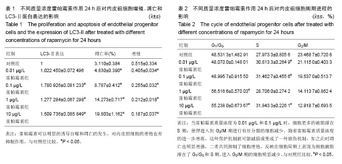
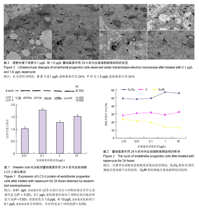
背景:曾有报道雷帕霉素可以对内皮祖细胞的增殖能力、迁移能力、黏附能力产生影响,但是没有提到自噬在其中所起到的不可忽视的作用,以及自噬与凋亡之间的相互关系。
目的:通过雷帕霉素激活自噬探讨自噬激活对大鼠内皮祖细胞增殖、凋亡和周期的影响。
方法:采用密度梯度离心法从骨髓获得单个核细胞,将其接种在人纤维连接蛋白包被的培养板上,培养7 d后收集贴壁细胞,即内皮祖细胞。加入不同质量浓度雷帕霉素(0.01,0.1,1,10 μg/ L)分别培养24 h。Western blot检测LC3-Ⅱ蛋白表达监测自噬的诱导情况,流式细胞仪检测细胞凋亡和细胞周期进程的变化,MTT比色法观察其增殖能力的变化,同时在透射电镜下观察其超微结构的变化。
结果与结论:雷帕霉素质量浓度为0.01 μg/L时,内皮祖细胞的LC3-Ⅱ蛋白表达与对照组相比并没有明显的增高,当质量浓度为0.1 μg/L时LC3-Ⅱ蛋白表达处在一个较高的水平,质量浓度为1 μg/L和10 μg/L时LC3-Ⅱ蛋白表达虽然也高于对照组,但却明显低于0.1 μg/L时,据此推断雷帕霉素在质量浓度为0.1 μg/L时自噬尤为活跃。内皮祖细胞的凋亡率呈现随着雷帕霉素质量浓度的升高而增加的趋势,增殖率呈现随雷帕霉质量浓度增加而降低的趋势。结果说明雷帕霉素激活自噬后能够促进细胞的凋亡,明显改变细胞的周期进程,抑制内皮祖细胞的增殖。


中国组织工程研究杂志出版内容重点:干细胞;骨髓干细胞;造血干细胞;脂肪干细胞;肿瘤干细胞;胚胎干细胞;脐带脐血干细胞;干细胞诱导;干细胞分化;组织工程


全文链接:

关键词: 干细胞, 培养, 雷帕霉素, 内皮祖细胞, LC3-Ⅱ蛋白, 自噬, 细胞凋亡, 细胞增殖, 细胞周期, 国家自然科学基金

Abstract:

BACKGROUND: Previous studies have reported that rapamycin can affect the proliferation, migration and adhesion abilities of endothelial progenitor cells, but there is no report on the effect of autophagy, as well as the interaction between autophagy and apoptosis.    
OBJECTIVE: To observe the effect of rapamycin activated autophagy activation on the proliferation, apoptosis, and cycle of endothelial progenitor cells. 
METHODS: Density gradient centrifugation was used to obtain mononuclear cells from bone marrow, and the mononuclear cells were inoculated on human fibronectin-coated culture plate.Then after cultured for 7 days the adherent cells collected were the endothelial progenitor cells. Different concentrations of rapamycin (0.01, 0.1, 1 and 10 μg/L) were added and cultured for 24 hours. Western blot was used to detect the LC3-II protein expression and monitor the induction of autophagy, flow cytometry was used to observe the cell cycle progression and apoptosis changes, and methylthiazolyldiphenyl-tetrazolium bromide colorimetric assay was used to observe the proliferation ability. Meanwhile, the ultrastructural changes were observed under transmission electron microscope. 
RESULTS AND CONCLUSION: Compared with the control group, there was no significant increasing of LC3-II protein expression of endothelial progenitor cells in 0.01 μg/L rapamycin group, and the LC3-II protein expression was in the high level. The LC3-II protein expression in the 1 μg/L and 10 μg/L rapamycin groups was higher than that in the control group, but lower than that in the 0.01 μg/L rapamycin group, which indicated that autophagy was particularly active when the concentration of rapamycin was 0.01 μg/L. The apoptosis of endothelial progenitor cells was increased with the increasing of concentration of rapamycin, and the proliferation rate was decreased with the increasing of concentration of rapamycin. The results indicate that activation of autophagy by bapamycin can promote the cell apoptosis, change the cell cycle significantly, and can inhibit the proliferation of endothelial progenitor cells.


中国组织工程研究杂志出版内容重点:干细胞;骨髓干细胞;造血干细胞;脂肪干细胞;肿瘤干细胞;胚胎干细胞;脐带脐血干细胞;干细胞诱导;干细胞分化;组织工程


全文链接:

Key words: Sirolimus, Autophagy, Cell cycle, Apoptosis, Cell Proliferation

中图分类号: