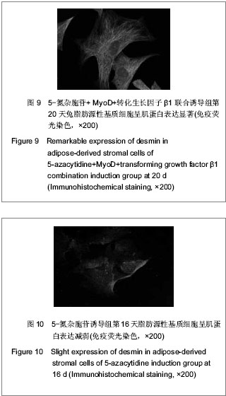
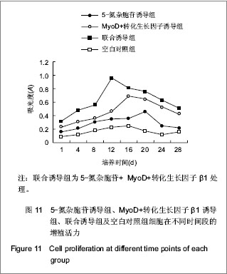
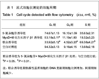
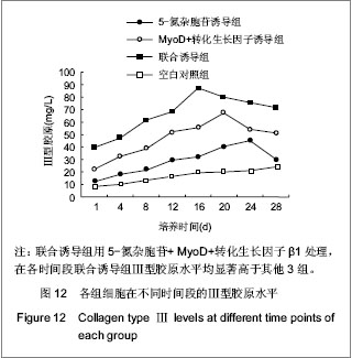
| [1] Hashimoto N, Murase T ,Kondo S, et al.Muscle reconstitution by muscle satellite cell descendants with stem cell-like properties.Development.2004;131(21):5481-5490.[2] Buckinghanl M.Skeletal muscle progenitor cells and the role of Pax genes.C R Biol.2007;330(6)530-533.[3] Konning M, Werker PM, Van Luyn MJ, et al. A global downregulation of microRNAs occurs in human quiescent satellite cells during myogenesis. Differentiation.2012; 84(4): 314-321.[4] Perrucho MH, Ecolan P, Sorensen IL, et al. In vitro characterization of proliferation and differentiation of pig satellite cells. Differentiation.2012;84(4):322-329.[5] Le Grand F, Grifone R, Mourikis P, et al. Six1 regulates stem cell repair potential and self-renewal during skeletal muscle regenerarion. J Cell Biol.2012;198(5):815-832.[6] Brohl D, Vasyutina E, Czajkowski MT, et al. Colonization of the satellite cell niche by skeletal muscle progenitor cells depends on notch signals. Dev Cell.2012;23(3):469-481.[7] 刘杰,傅强.Beagle犬上皮细胞原代培养鉴定及脂肪干细胞诱导成肌的实验研究[J].中华临床医师杂志,2011,5(10):2864- 2871.[8] 刘静,糜若然.组织工程学技术治疗女性张力性尿失禁研究进展[J].中国妇幼保健,2010,25(2):278-280.[9] Mitterberger M, Pinggera GM, Marksteiner R,et al. Adult stem cell therapy of female stress urinary incontinence. Eur Urol. 2008;53(1):169-175.[10] Xu Y, Fu W, Li G, et al. Autologous urothelial cells transplantation onto a prefaricated capsular stent for tissue engineered ureteral reconstruction. J Mater Sci Mater Med. 2012;23(4):1119-1128.[11] Chun SY, Kim HT, Lee JS, et al. Characterization of urine-derived cells frm upper urinary tract in patients with bladder cancer. Urology.2012;79(5):1-7.[12] Zhang J, Gu GL, Liu GH, et al. Ureteral reconstruction using autologous tubular grafts for the management of ureteral strictures and defects: an experimental study. Urol Int.2012; 88(1):60-65.[13] Chen BS, Xie H, Zhang SL, et al. Tissue engineering of bladder using vascular endothelial growth factor gene-modified endothelial progenitor cells. Int J Artif Organs. 2011;34(12):1137-1146.[14] Basu J, Jayo MJ, Ilagan RM, et al. Regeneration of native-like neo-urinary tissue from nonbladder cell sources. Tissue Eng Part A.2012;18(9):1025-1034.[15] Fu Q, Cao YL. Tissue engineering and stem cell application of urethroplasty;from bench to bedside. Urology.2012;79(2): 246-253.[16] Davis NF, Mooney R, Piterina AV, et al. Construction and evaluation of urinary bladder bioreactor for urologic tissue-engineering purposes. Urology.2011;78(9):954-960.[17] Subramaniam R, Hinley J, Stahlschmidt J, et al. Tissue engineering potential of urothelial cells from diseased bladders.J Urol.2011;186(5):2014-2020.[18] Kim YT, Kim DK, Jankowski RJ,et al. Human muscle-derived cell injection in a rat model of stress urinary incontinence. Muscle Nerve.2008;36(3):391-393.[19] Kwon D, Kim Y, Pruchnic R,et al. Periurethral cellular injection: comparison of muscle-derived progenitor cells and fibroblasts with regard to efficacy and tissue contractility in an animal model of stress urinary incontinence. Urology.2006;68(2): 449-454.[20] Yu J,Li Y,Li M,et al.Oxidized low density lipoprotein induced transdifferentiation of bone marrow-derived smooth muscle-like cells into foam-like cells in vitro. Int J Exp Pathol. 2010;91(1):24-33.[21] Tian H, Bharadwaj S, Liu Y, et al. Differentiation of human bone marrow mesenchymal stem cells into bladder cells: potential for urological tissue engineering. Tissue Eng Part A.2010;16(5):1769-1779.[22] Choi YS, Vincent LG, Lee AR, et al. Mechanial derivation of functional myotubes from adipose-derived stem cells. Biomaterials.2012;33(8):2482-2491.[23] Arien-Zakay H, Lazarovici P, Nagler A. Tissue regeneration potential in human umbilical cord blood. Best Pract Res Clin Haematol.2010;23(2):291-303.[24] Hauser PV, De Fazio R, Bruno S, et al. Stem cells derived from human amniotic fluid contribute to acute kidney injury recovery. Am J Pathol.2010;177(4):2011-2021.[25] Longo UG, Loppini M, Berton A, et al. Stem cells from umbilical cord and placenta from musculoskeletal tissue engineering. Curr Stem Cell Res Ther.2012,7(4):272-281.[26] Zuk PA, Zhu M, Mizuno H, et al. Multilineage cells from human adipose tissue: implications for cell-based therapies. Tissue Eng.2001;7(2):211.[27] Cho HH, Kim YJ, Kim SJ, et al. Endogenous wnt signaling promotes proliferation and suppresses osteogenic differentiation in human adipose derived stromal cells. Tissue Eng.2006;12(1):111.[28] Lin CS. Advances in stem cell therapy for the lower urinary tract. World Journal Stem Cell.2010; 26(1):1-4.[29] Shiraishi T, Sumita Y, Wakamastu Y, et al. Formation of engineered bone with adipose stromal cells from buccal fat pad. J Dent Res.2012;9(6):592-597.[30] Man Y, Wang P,guo Y, et al. Angiogenic and osteogenic potential of platelet-rich plasma and adipose-derived stem cell laden alginate microspheres. Biomaterials.2012;33(34): 8802-8811.[31] Geng J, Liu G, Peng F, et al. Decorin promotes myogenic differentiation and mdx mice therapeutic effects after transplantation of rat adipose-derived stemm cells. Cytotherapy. 2012;14(7):877-886 |
.jpg)