中国组织工程研究 ›› 2013, Vol. 17 ›› Issue (2): 205-209.doi: 10.3969/j.issn.2095-4344.2013.02.004

• 骨组织构建 bone tissue construction • 上一篇    下一篇
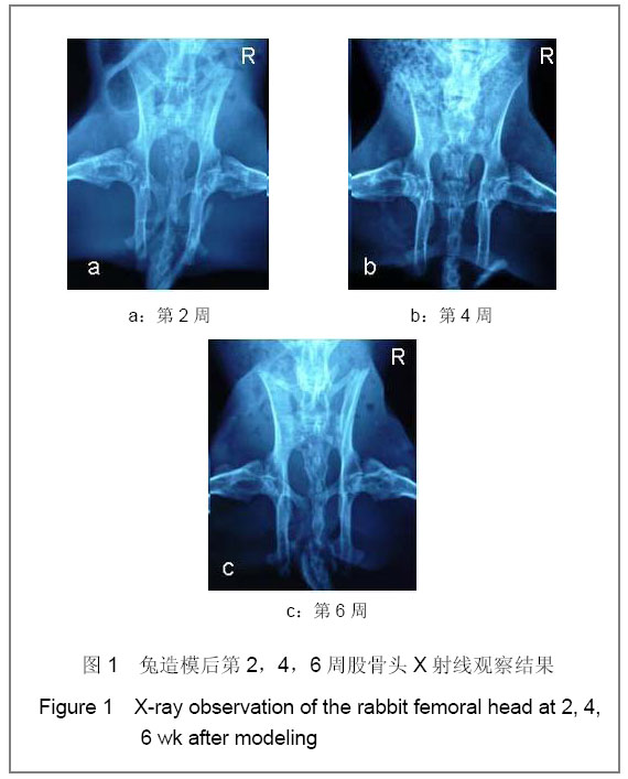
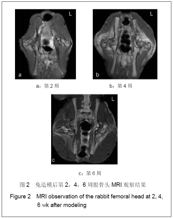
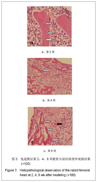
骨内注射无水乙醇建立兔股骨头坏死模型

陈 东1,华文彬1,叶树楠1,杨述华1,陈 超1,王小红1,禹 虔1,刘先哲1,索进平2,聂 磊2   

  1. 1 华中科技大学同济医学院附属协和医院骨科,湖北省武汉市  430022
    2 华中科技大学材料科学与工程学院,湖北省武汉市  430074
  • 收稿日期:2012-03-16 修回日期:2012-05-17 出版日期:2013-01-08 发布日期:2013-01-08
  • 通讯作者: 通信作者:杨述华,主任医师,教授,博士生导师,华中科技大学同济医学院附属协和医院骨科,湖北省武汉市 430022 shuhuayabc@vip.sina.com 并列通讯作者:叶树楠,博士,主治医师,华中科技大学同济医学院附属协和医院骨科,湖北省武汉市 430022 shunanye@gmail.com
  • 作者简介:陈东☆,男,1975年生,湖北省黄冈市人,华中科技大学同济医学院附属协和医院在读博士,主要从事股骨头坏死、骨组织工程研究。cd2408@163.com
  • 基金资助:

    中央高校基本科研业务费专项资金资助(2010JC029,2011JC042);国家自然科学基金资助项目(81171750,30973044)。

Intraosseous injection of pure alcohol induces necrosis of the femoral head in a rabbit

Chen Dong1, Hua Wen-bin1, Ye Shu-nan1, Yang Shu-hua1, Chen Chao1, Wang Xiao-hong1, Yu Qian1, Liu Xian-zhe1, Suo Jin-ping2, Nie Lei2   

  1. 1 Department of Orthopedics, Union Hospital, Tongji Medical College of Huazhong University of Science and Technology, Wuhan 430022, Hubei Province, China 2 Institute of Materials Science and Engineering, Huazhong University of Science and Technology, Wuhan 430074, Hubei Province, China
  • Received:2012-03-16 Revised:2012-05-17 Online:2013-01-08 Published:2013-01-08
  • Contact: Corresponding author: Yang Shu-hua, Chief physician, Doctoral supervisor, Professor, Department of Orthopedics, Union Hospital, Tongji Medical College of Huazhong University of Science and Technology, Wuhan 430022, Hubei Province, China shuhuayabc@vip.sina.com Corresponding author: Ye Shu-nan, Doctor, Attending physician, Department of Orthopedics, Union Hospital, Tongji Medical College of Huazhong University of Science and Technology, Wuhan 430022, Hubei Province, China shunanye@gmail.com
  • About author:Chen Dong☆, Studying for doctorate, Department of Orthopedics, Union Hospital, Tongji Medical College of Huazhong University of Science and Technology, Wuhan 430022, Hubei Province, China cd2408@163.com
  • Supported by:

    Supported by: the Fundamental Research Funds for the Central Universities, No. 2010JC029*, 2011JC042*; the National Natural Science Foundation of China, No. 81171750*, 30973044

摘要:

背景:目前还难以建立合适的早期股骨头坏死的动物模型。
目的:建立一个简单、标准和可靠的股骨头坏死动物模型用于实验研究。
方法:X射线透视下将新西兰兔右侧股骨头中心钻孔并注入无水乙醇,左侧不做处理做对照。经过2,4和6周麻醉下处死动物获取股骨头。
结果与结论:大体及X射线观察显示,造模侧股骨头第2周关节软骨颜色变暗、骨质密度不均匀;第6周,关节面有轻微凹陷,骨质低密度影较前进一步增大。MRI显示,造模侧股骨头第2周,T1加权股骨头负重区显示线样或不规则低信号,T2加权呈高信号;第6周,股骨头变性,软骨下骨折,关节面塌陷,新月体形成。对照侧第2,4,6周股骨头结果均常。组织病理学观察显示,造模侧股骨头第2周后,骨细胞核固缩、变性坏死。结果证实,2周后兔股骨头均发生了部分坏死,股骨头坏死的完整的外观形态,大体循环和关节软骨与人类早期股骨头坏死是相似的,提示实验建立了良好的股骨头坏死动物模型。

关键词: 组织构建, 骨组织构建, 股骨头坏死, 动物模型, 乙醇, 兔, X射线, MRI, 国家自然科学基金, 组织构建图片文章

Abstract:

BACKGROUND: Currently, it is difficult to prepare an animal model of early osteonecrosis of the femoral head (ONFH).
OBJECTIVE: To develop a simple and dependable animal model of ONFH that allows the evaluation of different therapeutic approaches.
METHODS: Pure alcohol was injected into the centre of the right femoral head in New Zealand white rabbits under X-ray fluoroscopy, and the left femoral head with no treatment served as controls. After 2, 4 and 6 weeks, the animals were killed under anesthesia and the femoral heads were harvested.
RESULTS AND CONCLUSION: Gross and X-ray observations indicated that the articular cartilage in the modeling side of the femoral head was dark in color and had the uneven bone density at the first two weeks after modeling. At 6 weeks, a slight depression was found on the articular surface, and the bone density shadow increased. MRI results showed that line or irregular T1-weighted signals and high T2-weighted signals were seen in the weight-bearing area of the femoral head at 2 weeks; femoral head degeneration, subchondral fracture, articular surface collapse, and crescent formation occurred at 6 weeks after modeling. In addition, there was no abnormality in the control side of the femoral head at 2, 4 and 6 weeks. Histopathological results showed karyopyknosis, degeneration and necrosis of the femoral head at 2 weeks after modeling. These findings verified that partial osteonecrosis was confirmed over a period of 2 weeks in all animals. The morphology of necrosis in combination with intact macrotexture and joint cartilage was similar to the features described in early ONFH in humans. We obtained a good efficacy and similarity animal model of the early stages of ONFH in humans. This model may be compatible to develop new therapeutic methods for ONFH.

Key words: tissue construction, bone tissue construction, femoral head necrosis, animal models, ethanol, rabbits, X-ray, MRI, National Natural Science Foundation of China, tissue construction photographs-containing paper

中图分类号: