[1] 刘晓东,张勉,王美青.咬合紊乱导致颞下颌关节髁突异常改建[J].中国实用口腔科杂志 2017,10(6):335-340.
[2] 李健,傅开元,冯海兰.颞下颌关节紊乱病的治疗[J].口腔颌面修复学杂志,2001,2(3):187-189.
[3] ZHANG J, JIAO K, ZHANG M, et al. Occlusal effects on longitudinal bone alterations of the temporomandibular joint. J Dent Res. 2013;92(3): 253-259.
[4] CAO Y, XIE QF, LI K, et al. Experimental occlusal interference induces long-term masticatory muscle hyperalgesia in rats. Pain. 2009;144(3): 287-293.
[5] THILANDER B, RUBIO G, PENA L, et al. Prevalence of temporomandibular dysfunction and its association with malocclusion in children and adolescents: an epidemiologic study related to specified stages of dental development. Angle Orthod. 2002;72(2):146-154.
[6] MCNAMARA JA Jr. Orthodontic treatment and temporomandibular disorders. Oral Surg Oral Med Oral Pathol Oral Radiol Endod. 1997; 83(1):107-117.
[7] LUTHER F. TMD and occlusion part I. Damned if we do? Occlusion: the interface of dentistry and orthodontics. Br Dent J. 2007;202(1):E2, 38-39.
[8] 李静,王旭霞,李涛,等.前方牵引反作用力对颞下颌关节区受力状况影响的有限元研究[J].临床口腔医学杂志,2013,29(9):519-523.
[9] 董瑞,王旭霞,张文娟,等.前牵引颏部反作用力对颞下颌关节影响的三维有限元分析[J].中华口腔医学杂志,2013,48(12):740-744.
[10] 杨辉,刘洪臣.不同方向颏兜力作用下颞下颌关节受力的三维有限元分析[J].口腔正畸学,1999,6(4):147-149.
[11] TANAKA E, TANNE K, SAKUDA M. A three-dimensional finite element model of the mandible including the TMJ and its application to stress analysis in the TMJ during clenching. Med Eng Phys. 1994;16(4): 316-322.
[12] FIELDS HW, SARVER DM, PROFFIT WR. Contemporary orthodontics.5nd ed.Mosby:Year Book,2013:462.
[13] GRANDORI F, MERLINI C, AMELOTTI C, et al. A mathematical model for the computation of the forces exerted by the facial orthopedic mask. Am J Orthod Dentofacial Orthop. 1992;101(5):441-448.
[14] BACCETTI T, ANTONINI A, FRANCHI L,et al. Glenoid fossa position in different facial types: a cephalometric study. Br J Orthod. 1997;24(1): 55-59.
[15] BUSCHANG PH, GANDINI JÚNIOR LG. Mandibular skeletal growth and modelling between 10 and 15 years of age. Eur J Orthod. 2002; 24(1):69-79.
[16] BUSCHANG PH, SANTOS-PINTO A. Condylar growth and glenoid fossa displacement during childhood and adolescence. Am J Orthod Dentofacial Orthop. 1998;113(4):437-442.
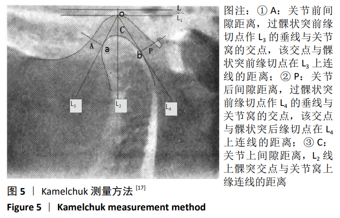
[17] KAMELCHUK LS, GRACE MG, Major PW. Post-imaging temporomandibular joint space analysis. Cranio. 1996;14(1):23-29.
[18] BJORK A. The nature of facial prognathism and its relation to normal occlusion of the teeth. Am J Orthod. 1951;37(2):106-124.
[19] KIM JH, VIANA MA, GRABER TM, et al. The effectiveness of protraction face mask therapy: a meta-analysis. Am J Orthod Dentofacial Orthop. 1999;115(6):675-685.
[20] 刘春艳,卢海燕,王雯,等.支架式与固定式前牵引矫治替牙期骨性Ⅲ类错牙合的比较[J].实用口腔医学杂志,2014,30(2):227-231.
[21] COPRAY JC, JANSEN HW, DUTERLOO HS. Growth and growth pressure of mandibular condylar and some primary cartilages of the rat in vitro. Am J Orthod Dentofacial Orthop. 1986;90(1):19-28.
[22] 王瑾,杨春.前牵引颏部反作用力对颞下颌关节应力及位移的影响[J].中国口腔颌面外科杂志,2015,13(3):216-219.
[23] 王瑞永,马绪臣,张万林,等.健康成年人颞下颌关节间隙锥形束计算机体层摄影术测量分析[J].北京大学学报(医学版), 2007, 39( 5):503-506.
[24] WEINBERG LA. Role of condylar position in TMJ dysfunction-pain syndrome. J Prosthet Dent. 1979;41(6):636-643.
[25] FARRAR WB, MCCARTY WL JR. Inferior joint space arthrography and characteristics of condylar paths in internal derangements of the TMJ. J Prosthet Dent. 1979;41(5):548-555.
[26] PULLINGER AG, SOLBERG WK, HOLLENDER L, et al. Tomographic analysis of mandibular condyle position in diagnostic subgroups of temporomandibular disorders. J Prosthet Dent. 1986;55(6):723-729.
[27] 汪传铎,王世平,魏民宪.110例颞下颌关节紊乱综合征薛氏位影像分析[J].中华口腔医学杂志,1990,25(2):117-119.
[28] 热比古丽·阿卜来提,迪丽努尔·阿吉,龚忠诚,等.锥形束CT测量颞下颌关节紊乱病患者颞下颌关节间隙的临床研究[J].中国美容医学杂志,2015,24(20):32-36.
[29] HU YK, YANG C, XIE QY. Changes in disc status in the reducing and nonreducing anterior disc displacement of temporomandibular joint: a longitudinal retrospective study. Sci Rep. 2016;6:34253.
[30] DE CLERCK H, NGUYEN T, DE PAULA LK, et al. Three-dimensional assessment of mandibular and glenoid fossa changes after bone-anchored Class III intermaxillary traction. Am J Orthod Dentofacial Orthop. 2012;142(1):25-31.
[31] YATABE M, GARIB D, FACO R, et al. Mandibular and glenoid fossa changes after bone-anchored maxillary protraction therapy in patients with UCLP: A 3-D preliminary assessment. Angle Orthod. 2017; 87(3): 423-431.
[32] MIMURA H, DEGUCHI T. Morphologic adaptation of temporomandibular joint after chincup therapy. Am J Orthod Dentofacial Orthop. 1996; 110(5):541-546. |