| [1] Ye YH,Chen K,Jin KK,et al.Progress on surgical treatment for femoral head-preservering in the precollapse stage of femoral head necrosis.Zhongguo Gu Shang. 2017;30(3):287-292.[2] Utsunomiya T,Motomura G,Ikemura S,et al.Effects of sclerotic changes on stress concentration in early-stage osteonecrosis: A patient-specific, 3D finite element analysis. J Orthop Res. 2018;36(12):3169-3177.[3] Issa K,Pivec R,Kapadia BH,et al.Osteonecrosis of the femoral head: the total hipreplacement solution.Bone Joint J. 2013; 95-B(11 Suppl A):46-50.[4] 朱旭日,杜斌,孙光权,等.髓芯减压打压植骨腓骨支撑术与头颈部开窗打压植骨术治疗早中期股骨头坏死疗效比较[J].中国骨与关节损伤杂志,2015,30(4):343-345.[5] 杜斌,孙鲁宁,袁滨,等.微创死骨清除打压植骨腓骨支撑配合中药补肾活血汤治疗早中期股骨头坏死的临床报道[J].中华关节外科杂志(电子版),2013,7(3):404-407.[6] Korompilias AV,Beris AE,Lykissas MG,et al. Femoral head osteonecrosis: why choose free vascularized fibula grafting. Microsurgery.2011;31(3):223-228.[7] 党莹,李月,李瑞玉,等.骨组织工程支架材料在骨缺损修复及3D打印技术中的应用[J].中国组织工程研究, 2017,21(14):2266-2273.[8] Šponer P,Ku?era T,Brtková J,et al.Comparative Study on the Application of Mesenchymal Stromal Cells Combined with Tricalcium Phosphate Scaffold into Femoral Bone Defects. Cell Transplant. 2018;27(10):1459-1468.[9] Bonazza V,Hajistilly C,Pate lD,et al.Growth Factors Release From Concentrated Growth Factors: Effect of β-Tricalcium Phosphate Addition. J Craniofac Surg.2018;29(8):2291-2295.[10] Wu JZ,Liu PC,Liu R. Icariin Restores Bone Structure and Strength in a Rat Model of Chronic High-Dose Alcohol-Induced Osteopenia. Cell Physiol Biochem.2018;46(4):1727-1736.[11] Jing X,Yin W,Tian H,et al. Icariin doped bioactive glasses seeded with rat adipose-derived stem cells to promote bone repair via enhanced osteogenic and angiogenic activities.Life Sci. 2018;202:52-60.[12] 薛鹏,杜斌,孙光权,等.可控释淫羊藿苷-β-磷酸三钙复合支架的制备[J].中国组织工程研究,2018,22(6):865-870.[13] The Ministry of Science and Technology of the People’s Republic of China. Guidance Suggestions for the Care and Use of Laboratory Animals.2006-09-30.中华人民共和国科学技术部.关于善待实验动物的指导性意见.2006-09-30.[14] 曹良权,杜斌,孙光权,等.不同剂量糖皮质激素联合马血清构建激素诱导型股骨头缺血性坏死兔模型[J].中国组织工程研究, 2017, 21(8):1229-1235.[15] Li D,Xie X,Kang P,et al.Percutaneously drilling through femoral head and neck fenestration combining with compacted autograft for early femoral head necrosis: A retrospective study. J Orthop Sci. 2017;22(6):1060-1065.[16] Zhu W,Zhao Y,Ma Q,et al.3D-printed porous titanium changed femoral head repair growth patterns: osteogenesis and vascularisation in porous titanium. J Mater Sci Mater Med. 2017;28(4):62.[17] Hu R,Lei P,Li B,et al.Real-time computerised tomography assisted porous tantalum implant in ARCO stage I-II non-traumatic osteonecrosis of the femoral head: minimum five-year follow up. Int Orthop. 2018;42(7):1535-1544.[18] Cheng Q,Tang JL,Gu JJ,et al.Total hip arthroplasty following failure of tantalum rod implantation for osteonecrosis of the femoral head with 5- to 10-year follow-up. BMC Musculoskelet Disord. 2018;19(1):289. [19] Hsieh TP,Sheu SY,Sun JS,et al.Icariin isolated from Epimedium pubescens regulates osteoblasts anabolism through BMP-2, SMAD4, andCbfa1expression. Phytomedicine. 2010;17(6): 414-423.[20] Shi W,Gao Y,Wang Y,et al.The flavonol glycoside icariin promotes bone formation in growing rats by activating the cAMP signaling pathway in primary cilia of osteoblasts. J Biol Chem. 2017;292(51):20883-20896.[21] Liu Y,Huang L,Hao B,et al.Use of an Osteoblast Overload Damage Model to Probe the Effect of Icariin on the Proliferation, Differentiation and Mineralization of MC3T3-E1 Cells through the Wnt/β-Catenin Signalling Pathway. Cell Physiol Biochem. 2017;41(4):1605-1615.[22] Ma XN,Ge BF,Chen KM,et al.Mechanisms of icariin in regulating bone formation of osteoblasts and bone resorption of osteoclasts.Zhongguo Yi Xue Ke Xue Yuan Xue Bao. 2013; 35(4):432-438.[23] Chung BH,Kim JD,Kim CK,et al. Icariin stimulates angiogenesis by activating the MEK/ERK- and PI3K/Akt/ eNOS-dependent signal pathways in human endothelial cells. Biochem Biophys Res Commun. 2008;376(2):404-408. |
.jpg)
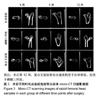
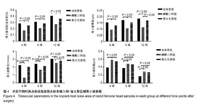
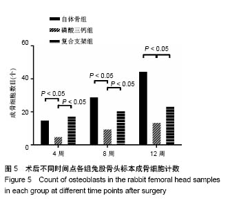
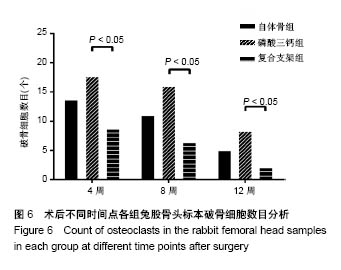
.jpg)
.jpg)
.jpg)
.jpg)