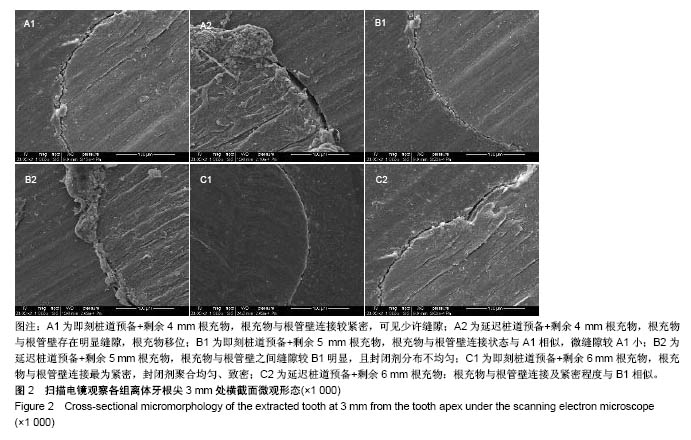
| [1] Siqueira JF Jr,Rocas IN,Ricucci D,et al. Causes and management of post-treatment apical periodontitis.Br Dent J. 2014;216(6):305-312.[2] Reyhani MF,Ghasemi N,Rahimi S,et al.Apical microleakage of AH Plus and MTA Fillapex sealers in association with immediate and delayed post space preparation: a bacterial leakage study.Minerva Stomatol.2015,64(3):129-134.[3] Metzger Z,Abramovitz R,Abramovitz I,et al.Correlation between remaining length of root canal fillings after immediate post space preparation and coronal leakage.J Endod. 2000; 26(12):724-728.[4] Pappen A,Bravo M,Gonzalez-Lopez S,et al.An in vitro study of coronal leakage after intraradicular preparation of cast-dowel space.J Prosthet Dent.2005;94(3):214-218.[5] Xu Q, Fan MW, Fan B, et al.A new quantitative method using glucose for analysis of endodontic leakage.Oral Surg Oral Med Oral Pathol Oral Radiol Endod.2005;99(1):107-111.[6] Rucucci D,Siqueira JF Jr.Recurrent apical periodontitis and late endodontic treatment failure related to coronal leakage: a case report.J Endod.2011;37(8):1171-1175.[7] Nikhil V,Jha P,Suri NK.Effect of methods of evaluation on sealing ability of mineral trioxide aggregate apical plug.J Consev Dent,2016,19(3):231-234.[8] 兰玉燕,黄海霞,范丽苑,等.不同材料充填根管后即刻与延迟桩道预备对根尖封闭性能的影响[J].中国组织工程研究, 2017, 21(10): 1483-1488.[9] Laslami K,Dhoum S,El Harchi A,et al. Relationship between the apical preparation diameter and the apical seal: an in vitro study.Int J Dent.2018;2018:2327854. [10] Moinzadeh AT,Mirmohammad H,Hensbergen IA,et al. The correlation between fluid transport and push-out strength in root canals filled with a methacrylate-based filling material.Int Endod J. 2015;48(2):193-198.[11] Ozkurt-Kayahan Z,Barut G,Ulusoy Z,et al.Influence of post space preparation on the apical leakage of calamus, single-cone and cold lateral condensation obturation techniques: a computerized fluid filtration study.J Prosthdont.2017.doi:10.1111/jopr.12623.[Epub ahead of print][12] Al-Maswary AA,Alhadainy HA,Al-Maweri SA.Coronal microleakage of the resilon and gutta-percha obtration materials with epiphany SE sealer: an in-vitro study.J Clin Diagn Res.2016;10(5):ZC39-42.[13] Mergud SS,Shah HH,Hiremath HT,et al.Effeict of post space preparation on the sealing ability of mineral trioxide aggregate and Gutta-percha: a bacterial leakage study.J Conserv Dent. 2015; 18(4):297-301.[14] Al-Madi EM,Al-Saleh SA,Al-Khudairy RI,et al.Influence of latrogenic gaps, cement type, and timie on microleakage pf cast posts using spectrophotometer and glucose filtration measurements.Int J Prosthodont.2018 Apr 6.doi:10.11607/ijp.5433.[Epub ahead of print][15] Kim SY,Ahn JS,Yi YA,et al.Quantitative microleakage analysis of endodontic temporary filling materials using a glucose penetration model.Acta Odontol Scand.2015;73(2):137-143.[16] Kececi AD,Kaya BU,Belli S.Corono‐apical leakage of various root filling materials using two different penetration models—a 3‐month study.J Biomed Mater Res B Appl Biomater. 2010,92(1):261-267.[17] Shemesh H,Wu MK,Wesselink P.Leakage along apical root fillings with and without smear layer using two different leakage models: a two‐month longitudinal ex vivo study.Int Endod J. 2006;39(12):968-976.[18] Padmanabhan P,Das J,Kumari RV,et al.Comparative evaluation of apical microleakage in immediate and delayed postspace preparation using four different root canal sealers: an in vitir study.J Conserv Dent.2017;20(2):86-90.[19] Zhou HM,Shen Y,Zheng W,et al.Physical propeties of 5 root canal sealers. J Endod. 2013; 39(10):1281-1286.[20] Solano F,Hartwell G,Appelstein C.Comparison of apical leakage between immediate versus delayed post space preparation using AH Plus sealer.J Endod. 2005;31(10): 752-754.[21] Dhaded N,Dhaded S,Patil C,et al.The effect of time of post space preparation on the seal and adaption of resilon-epithany Se and gutta-percha-AH Plus sealer-An SEM study.J Clin Diagn Res.2014;8(1):217-220.[22] Kim HR,Kim YK,Kwon TY.Post space preparation timing of root canals sealed with AH Plus sealer.Restor Dent Endod. 2017;42(1):27-33.[23] Su D,Hu X,Wang D,et al.Semiconductor laser irradiation improves root canal sealing during routine root canal therapy. PloS One.2017;12(9):e0186612.[24] 冯新颜,高承志.桩道超声清洗对两种根管封闭剂根尖微渗漏的影响[J].中国组织工程研究,2017,21(2):254-259.[25] Pusinanti L,Rubini R,Pellati A.A simplified post preparation technique after Thermafil obturation: evaluation of apical microleakage and presence of voids using methylene blue dye penetration.Ann Stomatol(Roma).2013;4(2):184-190.[26] Grecca FS,Rosa AR,Gomes MS,et al.Effect of timing and method of post space preparation on sealing ability of remaining root filling material: in vitro microbiological study.J Cant Dent Assoc.2009;75(8):583.[27] Jalalzadeh SM,Mamavi A,Abedi H,et al. Bacterial microleakage and post space timing for two endodontic sealers: an in vitro study.J Mass Dent Soc.2010;59(2):34-37.[28] Wang Z,Shen Y,Haapasalo M.Dentin extends the antibacterial effect of endodontic sealers against Enterococcus faecalis.J Endod.2014;4(4):505-508.[29] Ricketts D,Tait C,Higgins A.Tooth preparation for post-retained restoration.Br Dent J.2005;43(9):775-781.[30] 曲贞珍,李晓光,王青.桩道预备后剩余根管充填物的根尖封闭作用评价[J].上海口腔医学,2016,25(5):560-565.[31] Schuartz RS,Robbins JW.Post placement and restoration of endodontically treated teeth: a literature review.J Endod. 2004; 30(5):289-301.[32] 王洁琪,郑美华.桩核修复微渗漏影响因素研究进展[J].口腔颌面修复学杂志,2015,16(4):250-252. |
.jpg)


.jpg)