中国组织工程研究 ›› 2020, Vol. 24 ›› Issue (4): 517-523.doi: 10.3969/j.issn.2095-4344.2223

• 组织工程口腔材料 tissue-engineered oral materials • 上一篇    下一篇

不同玷污层对4种树脂粘接系统粘接强度及粘接耐久性的影响

郑适泽1,2,李  轲1,陈  玥1,鄢晓媛1,战德松3,付佳乐3   

  1. 1中国医科大学口腔医学院,辽宁省沈阳市  110002;2吉林大学口腔医学院,吉林省长春市  130021;3中国医科大学口腔医学院口腔材料学教研室,中国医科大学附属口腔医院修复二科,辽宁省沈阳市  110002
  • 收稿日期:2019-06-05 修回日期:2019-06-25 接受日期:2019-09-07 出版日期:2020-02-08 发布日期:2019-12-30
  • 通讯作者: 付佳乐,口腔医学博士,讲师,主治医师,中国医科大学口腔医学院,中国医科大学附属口腔医院,辽宁省沈阳市 110002
  • 作者简介:郑适泽,女,1996年生,吉林省长春市人,汉族,吉林大学口腔医学院在读硕士,主要从事粘接修复研究。
  • 基金资助:
    2018年辽宁省大学生创新创业训练计划资助项目(1111251703);中国医科大学新教师基金项目(XZR20160015)

Effect of different smear layers on the bonding performance and durability of four adhesive systems

Zheng Shize1, 2, Li Ke1, Chen Yue1, Yan Xiaoyuan1, Zhan Desong3, Fu Jiale3   

  1. 1School of Stomatology, China Medical University, Shenyang 110002, Liaoning Province, China; 2School of Stomatology, Jilin University, Changchun 130021, Jilin Province, China; 3Department of Dental Materials, School of Stomatology, China Medical University, The Second Department of Prosthodontics, Hospital of Stomatology, China Medical University, Shenyang 110002, Liaoning Province, China
  • Received:2019-06-05 Revised:2019-06-25 Accepted:2019-09-07 Online:2020-02-08 Published:2019-12-30
  • Contact: Fu Jiale, PhD, Lecturer, Attending Doctor, Department of Dental Materials, School of Stomatology, China Medical University, The Second Department of Prosthodontics, Hospital of Stomatology, China Medical University, Shenyang 110002, Liaoning Province, China
  • About author:Zheng Shize, Master candidate, School of Stomatology, China Medical University, Shenyang 110002, Liaoning Province, China; School of Stomatology, Jilin University, Changchun 130021, Jilin Province, China
  • Supported by:
    the Financial Aid Program on Innovation and Entrepreneurship for College Students in Liaoning Province in 2018, No. 1111251703; the New Teacher Foundation of China Medical University, No. XZR20160015

摘要:

文题释义:
玷污层:牙体预备时由于车针的高速切割和挤压,牙本质表面形成厚1-5 μm的玷污层,它由无机物碎屑和凝固的胶原纤维碎屑组成。切割碎屑深入牙本质小管口形成管塞。磷酸酸蚀处理能够去除牙本质表面的玷污层及污物,而自酸蚀粘接剂则因为酸性较弱仅能对牙本质表面的玷污层及杂质进行部分溶解,并最终将剩余的玷污层包含在内形成混合粘接层。
甲基丙烯酰癸氧基二氢磷酸酯(10-MDP):是一种性能优良的酸性功能单体,与羟基磷灰石反应生成的难溶性钙盐形成“纳米层状结构”,强烈吸附于牙本质表面,这一特点被认为能够增强粘接性能,并且对胶原纤维有保护作用,长期观察发现其可以提高粘接剂的耐久性。

背景:当用旋转式或手动器械处理牙表面时,在牙釉质和牙本质上由切割和磨损产生的碎片形成了玷污层。玷污层是影响牙和修复材料之间粘接的重要因素,明确玷污层对不同种类粘接剂的影响对口腔医生的临床选择与正确使用具有重要的临床意义。

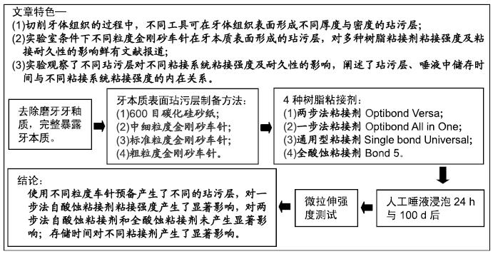
目的:评价使用不同金刚砂车针预备的试件在人工唾液中存储24 h与100 d后,对4种粘接剂与牙本质粘接强度的影响。

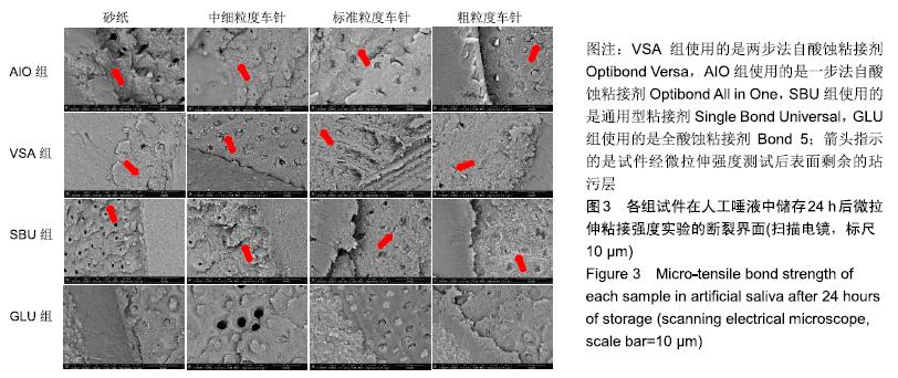
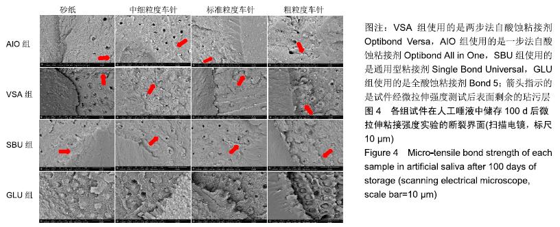
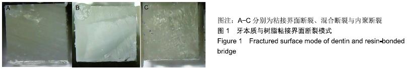
方法:获取离体人磨牙80颗,分别进行600号碳化硅砂纸(对照)、中细粒度金刚砂车针、标准粒度金刚砂车针、粗粒度金刚砂车针4种牙本质表面处理,每种处理20颗;每种处理方法内分4个亚组,分别以两步法自酸蚀粘接剂Optibond Versa(VSA组)、一步法自酸蚀粘接剂Optibond All in One(AIO组)、通用型粘接剂Single Bond Universal(SBU组)及全酸蚀粘接剂Bond 5(GLU组)进行树脂粘接。粘接完成后,将试件置于人工唾液中37 ℃储存24 h及100 d,随后进行微拉伸强度测试,使用扫描电镜观察牙本质端断裂面,并对断裂模式进行分析。

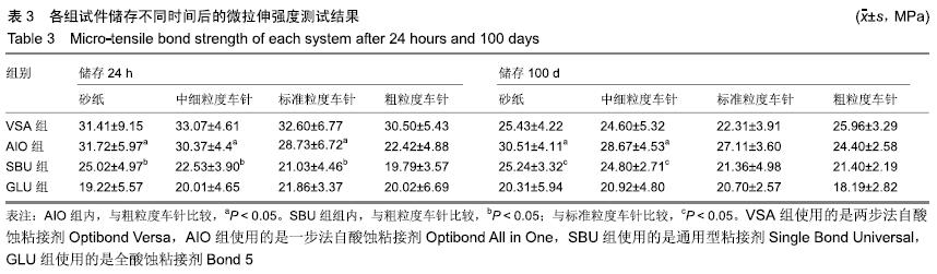
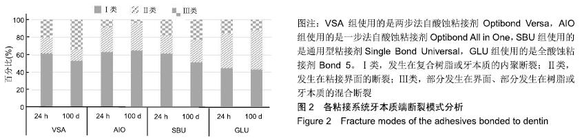
结果与结论:①储存24 h:使用两步法粘接剂或全酸蚀粘接剂的情况下,不同表面处理组间的粘接强度比较差异无显著性意义(P > 0.05);使用一步法粘接剂或通用型粘接剂的情况下,仅粗粒度车针组粘接强度低于砂纸组(P < 0.05)。使用砂纸或中细粒度车针的情况下,仅AIO组与VSA组粘接强度比较无差异;使用标准粒度车针的情况下,仅AIO组与VSA组、GLU组与SBU组粘接强度比较无差异(P > 0.05);使用粗粒度车针的情况下,VSA组与AIO组、GLU组、SBU组粘接强度比较差异均有显著性意义(P < 0.05),其余组间比较无差异(P > 0.05);②储存100 d:使用两步法粘接剂或全酸蚀粘接剂的情况下,不同表面处理组间的粘接强度比较差异无显著性意义(P > 0.05);使用一步法粘接剂的情况下,仅粗粒度车针组粘接强度低于砂纸组(P < 0.05);使用通用型粘接剂的情况下,仅标准粒度车针组、粗粒度车针组粘接强度低于砂纸组(P < 0.05)。使用砂纸或中细粒度车针的情况下,仅VSA组与SBU组粘接强度无差异(P > 0.05);使用标准粒度车针的情况下,仅SBU组与VSA组、GLU组与SBU组、GLU组与VSA组粘接强度无差异(P > 0.05);使用粗粒度车针的情况下,仅VSA组与AIO组粘接强度无差异(P > 0.05);③仅在使用两步法粘接剂的情况下,不同表面处理组100 d后的粘接强度与24 h比较差异有显著性意义(P < 0.05);④与浸泡24 h相比,VSA组、SBU组、GLU组浸泡100 d后的粘接界面断裂和混合断裂比例略有增加,其中VSA组变化最大;与其他组比较,GLU组浸泡24 h与100 d的粘接界面断裂和混合断裂模式比例均超过50%;⑤结果表明,使用不同粒度车针预备产生了不同的玷污层,对一步法自酸蚀粘接剂粘接强度产生了显著影响,对两步法自酸蚀粘接剂和全酸蚀粘接剂未产生显著影响;存储时间对不同粘接剂产生了显著影响。

ORCID: 0000-0002-1639-6498(郑适泽)

中国组织工程研究杂志出版内容重点:生物材料;骨生物材料; 口腔生物材料; 纳米材料; 缓释材料; 材料相容性;组织工程

关键词: 牙本质, 金刚砂车针, 玷污层, 通用型粘接剂, 微拉伸测试, 粘接强度, 扫描电镜, 断面观察

Abstract:

BACKGROUND: When the tooth surface is treated with a rotary or manual instrument, a smudge layer is formed on the enamel and dentin by debris generated by cutting and abrasion. The bonding interface between the adhesive and the dentin is considered as a weak part in the direct repair process. To clarify the effect of smear layer on different kinds of adhesive is significantly important for dentists to select and correctly use the adhesive in clinical treatment.

OBJECTIVE: To evaluate the effect of different diamond burs on the dentin bonding performance of four adhesive systems to dentin after 24 hours and 100 days of artificial saliva-storage.

METHODS: Adhesive systems were: (1) VSA (Optibond Versa, Kerr); (2) AIO (Optibond All in One, Kerr); (3) SBU (Single bond Universal, 3M); (4) GLU (Bond 5, Gluma,Heraeus). In present study, 80 extracted human molars were randomly divided into four groups and each group is divided into 4 subgroups. Dentin surfaces were prepared by: (1) 600-grit SiC-paper (control group); (2) super-fine diamond bur; (3) regular diamond bur; (4) coarse diamond bur. Bonding agent was applied according to each manufacturer’s instruction. After light-curing, dentin surfaces were built-up with resin composite (A2, CHARISMA, Heraeus). The micro-tensile bond strength was determined after 24 hours and 100 days of storage in artificial saliva at 37 °C. The fractured surfaces on dentin side were observed by scanning electron microscope.

RESULTS AND CONCLUSION: (1) Storage for 24 hours: there was no significant difference among groups under VSA and GLU. Under SBU and AIO, the bond strength in the coarse diamond bur group was significantly lower than that in the control group (P < 0.05). Using 600-grit SiC-paper or regular diamond bur, the bond strength showed no significant difference only between AIO and VSA groups. Using regular diamond bur, the bond strength showed no significant difference in the VSA group compared with the AIO, GLU and SBU groups (P > 0.05). Using coarse diamond bur, the bond strength in the VSA group had significant difference compared with the AIO, GLU and SBU groups (P < 0.05), and other groups had no significant difference (> 0.05). (2) Storage for 100 days, there was no significant difference among groups under VSA and GLU. Under SBU, the bond strength in the coarse diamond bur group was significantly lower than that in the control group (P < 0.05). Using 600-grit SiC-paper, the bond strength showed no significant difference only between VSA and SBU groups (P > 0.05). Using regular diamond bur, the bond strength showed no significant difference in the GLU group compared with the SBU, and VSA groups (> 0.05). Using coarse diamond bur, the bond strength had no significant difference between VSA and AIO groups (< 0.05). (3) Using VSA, the bond strength in each group at 100 days showed significant difference compared with that at 24 hours (P < 0.05). (4)Compared with immersed for 24 hours, the ratio of bond interface and combined crack in the VSA, SBU and GLU groups after immersed for 100 days was increased, especially the VSA group. Compared with the other groups, the ratio of bond interface and combined crack in the GLU group after immersed for 24 hours and 100 days both increased by 50%. (5) These results indicate that preparation by different burs produces different smear layers, which has significant effect to self-etching adhesive system and has no significant effect to total-etching adhesive system. Storage time makes effect on different adhesives.

Key words: dentin, diamond bur, smear layer, universal system, micro-tensile bond strength, bonding performance, scanning electrical microscope, fractured surface observation

中图分类号: