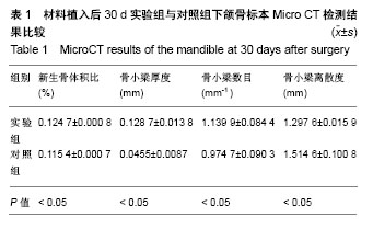
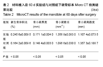
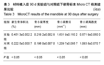
| [1] Sun J,Li YL,Chen LQ,et al.An experimental study on reconstruction of the condyle of the temporomandibular joint using free autogenous costal perichondrial grafts in rabbits. Oral Med Oral Pathol Oral Radiol Endodontol. 2010;109(5):649-796.[2] Shimada K,Tanaka H,Matsumoto T,et al.Cylindrical costal osteochondral autograft for reconstruction of large defects of the capitellum due to osteochondritis dissecans.J Bone Joint Surg Am. 2012;94(11):992-1002.[3] Mori Y,Saito T,Chang SH,et al.Identification of fibroblast growth factor-18 as a molecule to protect adult articular cartilage by gene expression profiling.J Biol Chem. 2014;289(14):10192.[4] Klammert U,Mueller TD,Hellmann TV,et al.GDF-5 can act as a context-dependent BMP-2antagonist.BMC Biol. 2015;13(1):1-18.[5] Seemann P,Mundlos S,Reissner C,et al.Bone morphogenetic protein 2 (BMP2) variants with reduced BMP antagonist sensitivity: WO,US 8828937 B2[P].2014.[6] Holly VL,Widen SA,Famulski JK,et al.Sfrp1a and Sfrp5 function as positive regulators of Wnt and BMP signaling during early retinal development.Dev Biol.2014;388(2):192.[7] Osses N,Henríquez JP.Bone morphogenetic protein signaling in vertebrate motor neurons and neuromuscular communication.Front Cell Neurosci.2015;8:453. [8] 周庆梅,孙健,李亚莉,等.生长因子缓释系统复合纳米支架材料修复犬下颌骨缺损[J].中国组织工程研究, 2013,17(21):3816-3822.[9] 安世昌,孙健,李亚莉,等.复合壳聚糖纳米微球聚乳酸-羟基乙酸/纳米羟基磷灰石缓释载体系统对蛋白的缓释作用[J].中国组织工程研究与临床康复,2011,15(25):4615-4618.[10] Vishnubalaji R,Yue S,Alfayez M,et al.Bone morphogenetic protein 2(BMP2) induces growth suppression and enhances chemosensitivity of human colon cancer cells.Cancer Cell Int. 2016;16(1):77.[11] Zhou N,Li Q,Lin X,et al.BMP2 induces chondrogenic differentiation, osteogenic differentiation and endochondral ossification in stem cells. Cell Tissue Res.2016;366(1):101-111.[12] Çak?r-Özkan N,E?ri S,Bekar E,et al.The Use of Sequential VEGF- and BMP2-Releasing Biodegradable Scaffolds in Rabbit Mandibular Defects.J Oral Maxillofac Surg. 2017;75(1):221.e1-221.e14. [13] John PS,Jeffery OH.The critical size defect as perimentalmodel for craniomandibulofacia Nonunions.Clini Orthop Rel Res. 1986;20(5): 299-230.[14] Lee JC,Min HJ,Park HJ,et al.Synovial membrane-derived mesenchymal stem cells supported by platelet-rich plasma can repair osteochondral defects in a rabbit model.Arthroscopy. 2013;29(6): 1034-1046.[15] 荣小芳,吴琳,杨晓东,等.兔下颌骨临界骨缺损人工材料植入实验动物模型的建立[J].中国医科大学学报, 2008,37(1):62-64.[16] 臧晓龙,孙健,李亚莉,等.3D生物打印构建聚乳酸羟基乙酸/纳米羟基磷灰石支架骨形态发生蛋白2缓释复合体的实验研究[J].中国组织工程研究, 2016,20(16):2405-2411.[17] The Ministry of Science and Technology of the People’s Republic of China. Guidance suggestion of caring laboratory animals.2006-09-30.[18] 朱明华,杨成民.植入性高分子材料的组织病理学观察[J].北京生物医学工程,1991,10(2):10217.[19] Tan W,Krishnaraj R,Desai TA.Evaluation of nanostructuredcomposite collagen-chitosan matrices for tissue engineering.Tissue Eng. 2001; 7(2):203-210.[20] Rosenberg L.Chemical basis for the histological use ofsafranin O in the study of articular cartilage.J Bone Joint Surg Am.1971;53(1):69-82.[21] Martyna K,Katarzyna W,Ma?gorzata LS,et al.Chitosan and composite microsphere-basedscaffold for bone tissue engineering: evaluation of tricalcium phosphate contentinfluence on physical and biological properties.J Nat Gas Sci Eng.2015;26(3):1-12.[22] Adnan H,Kailash CG,Inn-Kyu K,et al.PLGA/nHA hybrid nanofiber scaffold as a nanocargo carrier of insulin for accelerating bone tissue regeneration.Nanoscale Res Lett.2014;9(1):314-316.[23] Yang XZ,Li AS. Electrospun Hydroxyapatite/BMP-2 Grafted PLLA Nanofibers for Guided Bone Rebuilding Scaffold.Adv Mater Res.2015; 1095:322-325.[24] 许尧祥,李亚莉,陈立强,等.壳聚糖微球/纳米羟基磷灰石/聚乳酸-羟基乙酸复合支架制备及其蛋白缓释效果:与单纯纳米羟基磷灰石/聚乳酸-羟基乙酸支架、壳聚糖微球的比较[J].中国组织工程研究与临床康复, 2010, 14(3):452-456.[25] 刘峰,孙健,李亚莉,等.骨形态发生蛋白2壳聚糖纳米缓释载体的制备及性能测定[J].上海口腔医学, 2015,24(2):147-150.[26] S Dhivya S,Ajita J,Selvamurugan N.Metallic Nanomaterials for Bone Tissue Engineering.J Biomed Nanotechnol. 2015;11(10):1675-1700.[27] Labate GF,Baino F,Brovarone CV,et al.A biomimetic approach to 3D scaffold architectural characterization for veterinarian bone tissue engineering[C]// E-MRS Spring Meeting.2015.[28] Naga SM,El-Maghraby HF,Sayed M,et al.Highly porous scaffolds made of nanosized hydroxyapatite powder synthesized from eggshells.J Ceram Sci Technol. 2015;6(3):237-243.[29] Repanas A,Mavrilas D,Glasmacher B,et al.Blend electrospinning of biodegradable chitosan/ Polycaprolactone fibers as a process to create scaffolds for cardiovascular tissue engineering[C]// Esb.2015.[30] Huang J,Fu H,Wang ZY,et al.BMSCs-laden gelatin/sodium alginate/carboxymethylchitosan hydrogel for 3D bioprinting.Rsc Adv. 016;6(110):108423-108430.[31] Maji K,Dasgupta S,Pramanik K,et al.Preparation and Evaluation of Gelatin- Chitosan-Nanobioglass 3D Porous Scaffold for Bone Tissue Engineering.Int J Biomater. 2016;(4):183-187.[32] Dan Y,Liu O,Liu Y,et al.Development of Novel Biocomposite Scaffold of Chitosan-Gelatin/Nanohydroxyapatite for Potential Bone Tissue Engineering Applications.Nanoscale Res Lett. 2016;11(1):487.[33] Rahman MS,Akhtar N,Jami HMl,et al.TGF-b/BMP signaling and other molecular events:regulation of osteoblastogenesis and bone formation. Bone Res.2015;3(1):11-30.[34] Drosse I,Volkmer E,Capanna R,et al.Tissue engineering for bone defect healing: An update on a multi-component approach.Injury. 2008;39(2):S9-S20. |
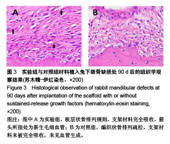
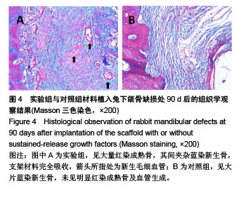
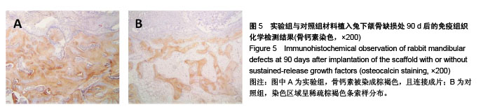
.jpg)
.jpg)
.jpg)