| [1]Zhu WZ, Hauch KD, Xu C, et al. Human embryonic stem cells and cardiac repair. Transplant Rev (Orlando). 2009;23(1):53-68.
[2]Hou D, Youssef EA, Brinton TJ, et al. Radiolabeled cell distribution after intramyocardial, intracoronary, and interstitial retrograde coronary venous delivery: implications for current clinical trials. Circulation. 2005;112(9 Suppl):I150-156.
[3]Suzuki K, Murtuza B, Fukushima S, et al.Targeted cell delivery into infarcted rat hearts by retrograde intracoronary infusion: distribution, dynamics, and influence on cardiac function. Circulation. 2004;110(11 Suppl 1):II225-230.[4] Silva GV, Litovsky S, Assad JA, et al. Mesenchymal stem cells differentiate into an endothelial phenotype, enhance vascular density, and improve heart function in a canine chronic ischemia model. Circulation. 2005;111(2):150-156.
[5]李鲁生,张涵,王成俊,等.骨髓间充质干细胞的分离方法和生物学特性[J].中国组织工程研究与临床康复,2010,14(10): 1869-1873.
[6]杨国凯,林明,许昌声,等.骨髓间充质干细胞移植对心力衰竭大鼠心功能的影响[J].中国心血管病研究,2009,7(6):458-461.
[7]Morigi M, Introna M, Imberti B, et al. Human bone marrow mesenchymal stem cells accelerate recovery of acute renal injury and prolong survival in mice. Stem Cells. 2008;26(8): 2075-2082.
[8]Shibata T, Naruse K, Kamiya H,et al. Transplantation of bone marrow-derived mesenchymal stem cells improves diabetic polyneuropathy in rats. Diabetes. 2008;57(11):3099-3107.
[9]Tang J, Wang J, Yang J, et al. Mesenchymal stem cells over-expressing SDF-1 promote angiogenesis and improve heart function in experimental myocardial infarction in rats. Eur J Cardiothorac Surg. 2009;36(4):644-650.
[10]Dowell JD, Rubart M, Pasumarthi KB, et al. Myocyte and myogenic stem cell transplantation in the heart. Cardiovasc Res. 2003;58(2):336-350.
[11]Mu Y, Cao G, Zeng Q, et al. Transplantation of induced bone marrow mesenchymal stem cells improves the cardiac function of rabbits with dilated cardiomyopathy via upregulation of vascular endothelial growth factor and its receptors.Exp Biol Med (Maywood). 2011;236(9):1100-1107.
[12]钟世根,凌智瑜,王志刚.超声微泡造影剂在血管新生中的应用研究进展[J].中华超声影像学杂志,2008,17(12):1088-1090.
[13]Nguyen BK, Maltais S, Perrault LP, et al. Improved function and myocardial repair of infarcted heart by intracoronary injection of mesenchymal stem cell-derived growth factors. J Cardiovasc Transl Res. 2010;3(5):547-558.
[14]Mansilla E, Marín GH, Drago H, et al. Bloodstream cells phenotypically identical to human mesenchymal bone marrow stem cells circulate in large amounts under the influence of acute large skin damage: new evidence for their use in regenerative medicine.Transplant Proc. 2006;38(3):967-969.
[15]Rüster B, Göttig S, Ludwig RJ, et al. Mesenchymal stem cells display coordinated rolling and adhesion behavior on endothelial cells. Blood. 2006;108(12):3938-3944.
[16]Ley K, Laudanna C, Cybulsky MI, et al. Getting to the site of inflammation: the leukocyte adhesion cascade updated. Nat Rev Immunol. 2007;7(9):678-689.
[17]Quattrocelli M, Cassano M, Crippa S, et al. Cell therapy strategies and improvements for muscular dystrophy. Cell Death Differ. 2010;17(8):1222-1229.
[18]江隆福,李恒栋.骨髓干细胞移植治疗急性前壁心肌梗死时间窗的选择[J].心脑血管病防治,2010,10(3):171-173.
[19]贾敏,卢沛琦,杨继要,等. 心肌梗死后不同时间移植骨髓间充质干细胞对心脏功能修复的作用[J].中国组织工程研究与临床康复,2007,11(33):6641-6644.
[20]Shen LH, Li Y, Chen J, et al. Therapeutic benefit of bone marrow stromal cells administered 1 month after stroke. J Cereb Blood Flow Metab. 2007;27(1):6-13.
[21]de Vasconcelos Dos Santos A, da Costa Reis J, Diaz Paredes B, et al. Therapeutic window for treatment of cortical ischemia with bone marrow-derived cells in rats. Brain Res. 2010;1306:149-158.
[22]Castillero E, Akashi H, Wang C, et al. Cardiac myostatin upregulation occurs immediately after myocardial ischemia and is involved in skeletal muscle activation of atrophy. Biochem Biophys Res Commun. 20150;457(1):106-111.
[23]Di Scipio F, Sprio AE, Folino A, et al. Injured cardiomyocytes promote dental pulp mesenchymal stem cell homing. Biochim Biophys Acta. 2014;1840(7):2152-2161.
[24]王广斌,付勤,杨礼庆,等.不同分离方法对骨髓间充质干细胞成软骨分化的影响[J].中国组织工程研究与临床康复,2008,12(38): 7577-7581.
[25]Li N, Yang H, Lu L, et al. Comparison of the labeling efficiency of BrdU, DiI and FISH labeling techniques in bone marrow stromal cells. Brain Res. 2008;1215:11-19.
[26]Fan X, Liu T, Liu Y, et al. Optimization of primary culture condition for mesenchymal stem cells derived from umbilical cord blood with factorial design. Biotechnol Prog. 2009;25(2): 499-507.
[27]Dvorakova J, Hruba A, Velebny V, et al. Isolation and characterization of mesenchymal stem cell population entrapped in bone marrow collection sets. Cell Biol Int. 2008;32(9):1116-1125.
[28]Fong EL, Chan CK, Goodman SB. Stem cell homing in musculoskeletal injury. Biomaterials. 2011;32(2):395-409.
[29]Yang CH, Sheu JJ, Tsai TH, et al. Effect of tacrolimus on myocardial infarction is associated with inflammation, ROS, MAP kinase and Akt pathways in mini-pigs. J Atheroscler Thromb. 2013;20(1):9-22.
[30]宋桂仙,李小荣,张凤祥,等. 猪心肌梗死后室性心律失常模型建立方法的比较[J].中国心脏起搏与心电生理杂志,2012,26(3): 246-249.
[31]Lichtenauer M, Schreiber C, Jung C, et al. Myocardial infarct size measurement using geometric angle calculation. Eur J Clin Invest. 2014;44(2):160-167.
[32]Cui X, Wang H, Guo H, et al. Transplantation of mesenchymal stem cells preconditioned with diazoxide, a mitochondrial ATP-sensitive potassium channel opener, promotes repair of myocardial infarction in rats. Tohoku J Exp Med. 2010;220(2): 139-147.
[33]Möbius C, Demuth C, Aigner T, et al. Evaluation of VEGF A expression and microvascular density as prognostic factors in extrahepatic cholangiocarcinoma. Eur J Surg Oncol. 2007; 33(8): 1025-1029.
[34]曹星宇,肖践明. 骨髓间充质干细胞归巢进展[J].临床医学,2013, 33(3): 111-114.
[35]Karp JM, Leng Teo GS. Mesenchymal stem cell homing: the devil is in the details. Cell Stem Cell. 2009;4(3):206-216.
[36]Shi M, Li J, Liao L, et al. Regulation of CXCR4 expression in human mesenchymal stem cells by cytokine treatment: role in homing efficiency in NOD/SCID mice.Haematologica. 2007; 92(7):897-904.
[37]Vandervelde S, van Luyn MJ, Tio RA, et al. Signaling factors in stem cell-mediated repair of infarcted myocardium. J Mol Cell Cardiol. 2005;39(2):363-376.
[38]Ma J, Ge J, Zhang S, et al. Time course of myocardial stromal cell-derived factor 1 expression and beneficial effects of intravenously administered bone marrow stem cells in rats with experimental myocardial infarction. Basic Res Cardiol. 2005;100(3):217-223.
[39]Askari AT, Unzek S, Popovic ZB, et al. Effect of stromal-cell-derived factor 1 on stem-cell homing and tissue regeneration in ischaemic cardiomyopathy. ancet. 2003;362 (9385):697-703.
[40]Li Y, Yu X, Lin S, et al. Insulin-like growth factor 1 enhances the migratory capacity of mesenchymal stem cells. Biochem Biophys Res Commun. 2007;356(3):780-784.
[41]Ma J, Ge J, Zhang S, et al. Time course of myocardial stromal cell-derived factor 1 expression and beneficial effects of intravenously administered bone marrow stem cells in rats with experimental myocardial infarction. Basic Res Cardiol. 2005;100(3):217-223.
[42]Kucia M, Dawn B, Hunt G, et al. Cells expressing early cardiac markers reside in the bone marrow and are mobilized into the peripheral blood after myocardial infarction. Circ Res. 2004;95(12):1191-1199.
[43]Bartunek J, Wijns W, Heyndrickx GR, et al. Timing of intracoronary bone-marrow-derived stem cell transplantation after ST-elevation myocardial infarction.Nat Clin Pract Cardiovasc Med. 2006;3 Suppl 1:S52-56.
[44]Hu X, Wang J, Chen J, et al. Optimal temporal delivery of bone marrow mesenchymal stem cells in rats with myocardial infarction. Eur J Cardiothorac Surg. 2007;31(3):438-443. |
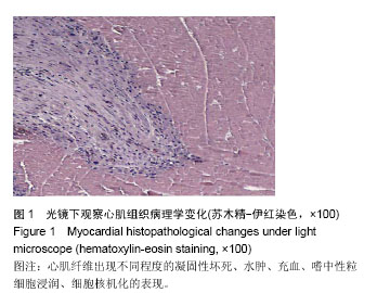
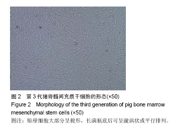
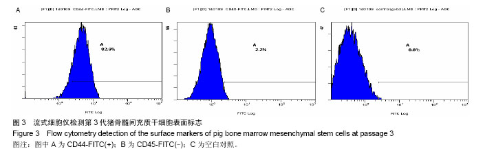
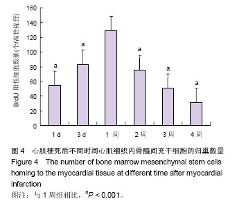
.jpg)