[1] GARCÍA-GARETA E, COATHUP MJ, BLUNN GW. Osteoinduction of bone grafting materials for bone repair and regeneration. Bone. 2015;81:112-121.
[2] HOFMANN GO, KIRSCHNER MH, WANGEMANN T, et al. Infections and immunological hazards of allogeneic bone transplantation. Arch Orthop Trauma Surg. 1995;114(3):159-166.
[3] 廖欣宇,王福科,王国梁.骨组织工程支架的进展与挑战[J].中国组织工程研究,2021, 25(28):4553-4560.
[4] FUKADA E, YASUDA I. On the piezoelectric effect of bone. J Phys Soc Jpn. 1957;12(10):1158-1162.
[5] MARTONOSI AN. Animal electricity, Ca2+ and muscle contraction. A brief history of muscle research. Acta Biochim Pol. 2000;47(3):493-516.
[6] HORAS K, ARNHOLDT J, STEINERT AF, et al. Acetabular defect classification in times of 3D imaging and patient-specific treatment protocols. Orthopade. 2017;46(2):168-178.
[7] SAIZ CJ, ESCOBAR HJ, GARZÓN-ALVARADO DA, et al. Electric and magnetic field devices for stimulation of biological tissues. J Vis Exp. 2021. doi: 10.3791/62111.
[8] LEVIN M. Bioelectric signaling: Reprogrammable circuits underlying embryogenesis, regeneration, and cancer. Cell. 2021;184(8):1971-1989.
[9] KEYNES RD. The ionic channels in excitable membranes. Ciba Found Symp. 1975;(31):191-203.
[10] DA SL, KUNDU SC, REIS L, et al. Electric phenomenon: a disregarded tool in tissue engineering and regenerative medicine. Trends Biotechnol. 2020;38(1):24-49.
[11] WIELAND DC, KRYWKA C, MICK E, et al. Investigation of the inverse piezoelectric effect of trabecular bone on a micrometer length scale using synchrotron radiation. Acta Biomater. 2015;25:339-346.
[12] DEVET T, JHIRAD A, PRAVATO L, et al. Bone bioelectricity and bone-cell response to electrical stimulation: a review. Crit Rev Biomed Eng. 2021;49(1):1-19.
[13] YANG M, BRACKENBURY WJ. Membrane potential and cancer progression. Front Physiol. 2013; 4:185.
[14] MYCIELSKA ME, DJAMGOZ MB. Cellular mechanisms of direct-current electric field effects: galvanotaxis and metastatic disease. J Cell Sci. 2004;117(Pt 9):1631-1639.
[15] ROBINSON KR. The responses of cells to electrical fields: a review. J Cell Biol. 1985;101(6):2023-2027.
[16] DIXON DT, GOMILLION CT. Conductive scaffolds for bone tissue engineering: current state and future outlook. J Funct Biomater. 2021;13(1):1.
[17] BASSETT CA, PAWLUK RJ, BECKER RO. Effects of electric currents on bone in vivo. Nature. 1964; 204:652-654.
[18] BEZANILLA F. The voltage sensor in voltage-dependent ion channels. Physiol Rev. 2000;80(2): 555-592.
[19] ZAYZAFOON M. Calcium/calmodulin signaling controls osteoblast growth and differentiation. J Cell Biochem. 2006;97(1):56-70.
[20] GRIFFIN M, BAYAT A. Electrical stimulation in bone healing: critical analysis by evaluating levels of evidence. Eplasty. 2011;11:e34.
[21] Li JK, Lin JC, Liu HC, et al. Comparison of ultrasound and electromagnetic field effects on osteoblast growth. Ultrasound Med Biol. 2006;32(5):769-775.
[22] MURILLO G, BLANQUER A, VARGAS-ESTEVEZ C, et al. Electromechanical nanogenerator-cell interaction modulates cell activity. Adv Mater. 2017. doi: 10.1002/adma.201605048.
[23] KIM IS, SONG JK, SONG YM, et al. Novel effect of biphasic electric current on in vitro osteogenesis and cytokine production in human mesenchymal stromal cells. Tissue Eng Part A. 2009;15(9):2411-2422.
[24] XU J, WANG W, CLARK CC, et al. Signal transduction in electrically stimulated articular chondrocytes involves translocation of extracellular calcium through voltage-gated channels. Osteoarthritis Cartilage. 2009;17(3):397-405.
[25] BRENNAN MÁ, LAYROLLE P, MOONEY DJ. Biomaterials functionalized with MSC secreted extracellular vesicles and soluble factors for tissue regeneration. Adv Funct Mater. 2020; 30(37):1909125.
[26] CIOMBOR DM, AARON RK. The role of electrical stimulation in bone repair. Foot Ankle Clin. 2005;10(4):579-593.
[27] ORR AW, HELMKE BP, BLACKMAN BR, et al. Mechanisms of mechanotransduction. Dev Cell. 2006;10(1):11-20.
[28] CHENG N, VAN HOOF H, BOCKX E, et al. The effects of electric currents on ATP generation, protein synthesis, and membrane transport of rat skin. Clin Orthop Relat Res. 1982;(171):264-272.
[29] ATKINSON SJ, HOSFORD MA, MOLITORIS BA. Mechanism of actin polymerization in cellular ATP depletion. J Biol Chem. 2004;279(7):5194-5199.
[30] TITUSHKIN I, CHO M. Regulation of cell cytoskeleton and membrane mechanics by electric field: role of linker proteins. Biophys J. 2009;96(2):717-728.
[31] SCHMELTER M, ATEGHANG B, HELMIG S, et al. Embryonic stem cells utilize reactive oxygen species as transducers of mechanical strain-induced cardiovascular differentiation. FASEB J. 2006. 20(8):1182-1184.
[32] DÍAZ-VEGAS A, CAMPOS CA, CONTRERAS-FERRAT A, et al. ROS production via P2Y1-PKC-NOX2 is triggered by extracellular ATP after electrical stimulation of skeletal muscle cells. PLoS One. 2015;10(6):e129882.
[33] ZHANG C, LIU W, CAO C, et al. Modulating surface potential by controlling the β phase content in poly (vinylidene fluoridetrifluoroethylene) membranes enhances bone regeneration. Adv Healthc Mater. 2018;7(11):e1701466.
[34] PEREDA AE. Electrical synapses and their functional interactions with chemical synapses. Nat Rev Neurosci. 2014;15(4):250-263.
[35] VENKATESH HS, MORISHITA W, GERAGHTY AC, et al. Electrical and synaptic integration of glioma into neural circuits. Nature. 2019;573(7775):539-545.
[36] KOTWAL A, SCHMIDT CE. Electrical stimulation alters protein adsorption and nerve cell interactions with electrically conducting biomaterials. Biomaterials. 2001;22(10):1055-1064.
[37] VACA-GONZÁLEZ JJ, GUEVARA JM, MONCAYO MA, et al. Biophysical stimuli: a review of electrical and mechanical stimulation in hyaline cartilage. Cartilage. 2019;10(2):157-172.
[38] SRIRUSSAMEE K, MOBINI S, CASSIDY NJ, et al. Direct electrical stimulation enhances osteogenesis by inducing Bmp2 and Spp1 expressions from macrophages and preosteoblasts. Biotechnol Bioeng. 2019;116(12):3421-3432.
[39] SHEIKH Z, HAMDAN N, IKEDA Y, et al. Natural graft tissues and synthetic biomaterials for periodontal and alveolar bone reconstructive applications: a review. Biomater Res. 2017;21:9.
[40] MONDSCHEIN RJ, KANITKAR A, WILLIAMS CB, et al. Polymer structure-property requirements for stereolithographic 3D printing of soft tissue engineering scaffolds. Biomaterials. 2017;140: 170-188.
[41] COSTA BC, TOKUHARA CK, ROCHA LA, et al. Vanadium ionic species from degradation of Ti-6Al-4V metallic implants: in vitro cytotoxicity and speciation evaluation. Mater Sci Eng C Mater Biol Appl. 2019;96:730-739.
[42] FROST HM. A 2003 update of bone physiology and Wolff’s Law for clinicians. Angle Orthod. 2004;74(1):3-15.
[43] HIEDA J, NIINOMI M, NAKAI M, et al. Adhesive strength of medical polymer on anodic oxide nanostructures fabricated on biomedical β-type titanium alloy. Mater Sci Eng C Mater Biol Appl. 2014;36:244-251.
[44] 杨铁威,刘欣伟,柳云恩,等.钛铌锆钽合金可促进兔肱骨干横骨折的愈合[J].中国组织工程研究,2014,18(52):8382-8386.
[45] WU H, DONG H, TANG Z, et al. Electrical stimulation of piezoelectric BaTiO3 coated Ti6Al4V scaffolds promotes anti-inflammatory polarization of macrophages and bone repair via MAPK/JNK inhibition and OXPHOS activation. Biomaterials. 2023;293:121990.
[46] XUE W, DU J, LI Q, et al. Preparation, properties, and application of graphene-based materials in tissue engineering scaffolds. Tissue Eng Part B Rev. 2022;28(5):1121-1136.
[47] WANG Q, CHU Y, HE J, et al. A graded graphene oxide-hydroxyapatite/silk fibroin biomimetic scaffold for bone tissue engineering. Mater Sci Eng C Mater Biol Appl. 2017;80:232-242.
[48] AIDUN A, SAFAEI FA, MOHARRAMI M, et al. Graphene oxide incorporated polycaprolactone/chitosan/collagen electrospun scaffold: enhanced osteogenic properties for bone tissue engineering. Artif Organs. 2019;43(10):E264-E281.
[49] BELAID H, NAGARAJAN S, TEYSSIER C, et al. Development of new biocompatible 3D printed graphene oxide-based scaffolds. Mater Sci Eng C Mater Biol Appl. 2020;110:110595.
[50] 陈琳.还原氧化石墨烯表面修饰硅酸钙生物陶瓷的制备及骨再生修复性能的研究[D].绵阳:西南科技大学,2019.
[51] CHLANDA A, WALEJEWSKA E, KOWIORSKI K, et al. Investigation into morphological and electromechanical surface properties of reduced-graphene-oxide-loaded composite fibers for bone tissue engineering applications: a comprehensive nanoscale study using atomic force microscopy approach. Micron. 2021;146:103072.
[52] AAMODT JM, GRAINGER DW. Extracellular matrix-based biomaterial scaffolds and the host response. Biomaterials. 2016;86:68-82.
[53] ZHAO C, XIAO Y, LING S, et al. Structure of collagen. Methods Mol Biol. 2021;2347:17-25.
[54] ZAREI M, SAMIMI A, KHORRAM M, et al. Fabrication and characterization of conductive polypyrrole/chitosan/collagen electrospun nanofiber scaffold for tissue engineering application. Int J Biol Macromol. 2021;168:175-186.
[55] COLLINS MN, BIRKINSHAW C. Hyaluronic acid based scaffolds for tissue engineering--a review. Carbohydr Polym. 2013;92(2):1262-1279.
[56] OHBA S, WANG W, ITOH S, et al. Efficacy of platelet-rich plasma gel and hyaluronan hydrogel as carriers of electrically polarized hydroxyapatite microgranules for accelerating bone formation. J Biomed Mater Res A. 2012;100(11):3167-3176.
[57] GUO L, LIANG Z, YANG L, et al. The role of natural polymers in bone tissue engineering. J Control Release. 2021;338:571-582.
[58] SHAABANI A, SEDGHI R. Preparation of chitosan biguanidine/PANI-containing self-healing semi-conductive waterborne scaffolds for bone tissue engineering. Carbohydr Polym. 2021; 264:118045.
[59] MCCULLEN SD, MCQUILLING JP, GROSSFELD RM, et al. Application of low-frequency alternating current electric fields via interdigitated electrodes: effects on cellular viability, cytoplasmic calcium, and osteogenic differentiation of human adipose-derived stem cells. Tissue Eng Part C Methods. 2010;16(6):1377-1386.
[60] SUN S, TITUSHKIN I, CHO M. Regulation of mesenchymal stem cell adhesion and orientation in 3D collagen scaffold by electrical stimulus. Bioelectrochemistry. 2006;69(2):133-141.
[61] TOPSAKAL A, UZUN M, UGAR G, et al. Development of Amoxicillin-Loaded Electrospun Polyurethane/Chitosan/ $\beta$ -Tricalcium Phosphate Scaffold for Bone Tissue Regeneration. IEEE Trans Nanobioscience. 2018;17(3):321-328.
[62] POURJAVADI A, DOROUDIAN M, AHADPOUR A, et al. Injectable chitosan/κ-carrageenan hydrogel designed with au nanoparticles: a conductive scaffold for tissue engineering demands. Int J Biol Macromol. 2019;126:310-317.
[63] ZHOU C, YE X, FAN Y, et al. Biomimetic fabrication of a three-level hierarchical calcium phosphate/collagen/hydroxyapatite scaffold for bone tissue engineering. Biofabrication. 2014; 6(3):35013.
[64] KIM DY, KWON DY, KWON JS, et al. Stimuli-responsive injectable in situ-forming hydrogels for regenerative medicines. Polym Rev. 2015;55(3):407-452.
[65] JING Y, QUAN C, LIU B, et al. A mini review on the functional biomaterials based on poly (lactic acid) stereocomplex. Polym Rev. 2016;56(2):262-286.
[66] LIN YJ, HUANG CC, WAN WL, et al. Recent advances in CO(2) bubble-generating carrier systems for localized controlled release. Biomaterials. 2017;133:154-164.
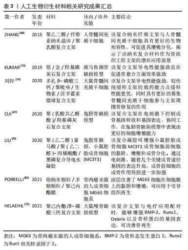
[67] CUI L, ZHANG J, ZOU J, et al. Electroactive composite scaffold with locally expressed osteoinductive factor for synergistic bone repair upon electrical stimulation. Biomaterials. 2020;230:119617.
[68] ZHANG C, SALICK MR, CORDIE TM, et al. Incorporation of poly(ethylene glycol) grafted cellulose nanocrystals in poly(lactic acid) electrospun nanocomposite fibers as potential scaffolds for bone tissue engineering. Mater Sci Eng C Mater Biol Appl. 2015;49:463-471.
[69] PORRELLI D, MARDIROSSIAN M, MUSCIACCHIO L, et al. Antibacterial electrospun polycaprolactone membranes coated with polysaccharides and silver nanoparticles for guided bone and tissue regeneration. ACS Appl Mater Interfaces. 2021;13(15):17255-17267.
[70] LIU X, GEORGE MN, LI L, et al. Injectable electrical conductive and phosphate releasing gel with two-dimensional black phosphorus and carbon nanotubes for bone tissue engineering. ACS Biomater Sci Eng. 2020;6(8):4653-4665.
[71] PALIERSE E, HÉLARY C, KRAFFT JM, et al. Baicalein-modified hydroxyapatite nanoparticles and coatings with antibacterial and antioxidant properties. Mater Sci Eng C Mater Biol Appl. 2021;118:111537.
[72] WANG W, ITOH S, TANAKA Y, et al. Comparison of enhancement of bone ingrowth into hydroxyapatite ceramics with highly and poorly interconnected pores by electrical polarization. Acta Biomater. 2009;5(8):3132-3140.
[73] KUMAR VB, KHAJURIA DK, KARASIK D, et al. Silver and gold doped hydroxyapatite nanocomposites for enhanced bone regeneration. Biomed Mater. 2019;14(5):55002.
[74] 刘锌,杜斌,孙光权,等.多孔β磷酸三钙-聚吡咯-生物素-淫羊藿素微球复合支架促进骨髓间充质干细胞的募集[J].中国组织工程研究,2020,24(34):5532-5537.
[75] HELAEHIL JV, LOURENÇO CB, HUANG B, et al. In vivo investigation of polymer-ceramic PCL/HA and PCL/β-TCP 3D composite scaffolds and electrical stimulation for bone regeneration. Polymers (Basel). 2021;14(1):65.
[76] DOROZHKIN SV. Bioceramics of calcium orthophosphates. Biomaterials. 2010;31(7):1465-1485.
[77] MATA D, OLIVEIRA FJ, NETO MA, et al. Smart electroconductive bioactive ceramics to promote in situ electrostimulation of bone. J Mater Chem B. 2015;3(9):1831-1845.
[78] JIANG N, GUO Z, SUN D, et al. Exploring the mechanism behind improved osteointegration of phosphorylated titanium implants with hierarchically structured topography. Colloids Surf B Biointerfaces. 2019;184:110520.
[79] FOROUTAN T, NAZEMI N, TAVANA M, et al. Suspended graphene oxide nanoparticle for accelerated multilayer osteoblast attachment. J Biomed Mater Res A. 2018;106(1):293-303.
[80] PAWELEC KM, BEST SM, CAMERON RE. Collagen: a network for regenerative medicine. J Mater Chem B. 2016;4(40):6484-6496.
[81] HANSON SE, BENTZ ML, HEMATTI P. Mesenchymal stem cell therapy for nonhealing cutaneous wounds. Plast Reconstr Surg. 2010;125(2):510-516.
[82] SHIGEEDA W, SHIBAZAKI M, YASUHIRA S, et al. Hyaluronic acid enhances cell migration and invasion via the YAP1/TAZ-RHAMM axis in malignant pleural mesothelioma. Oncotarget. 2017; 8(55):93729-93740.
[83] STELLAVATO A, PIROZZI A, Diana P, et al. Hyaluronic acid and chondroitin sulfate, alone or in combination, efficiently counteract induced bladder cell damage and inflammation. PLoS One. 2019,14(6):e218475.
[84] 贺庆,敖强,修波,等.关于壳聚糖材料生物相容性机制的新观点(英文)[J].中国组织工程研究与临床康复,2007,11(35):7110-7112.
[85] 李浩宇,张伟,冯凯,等.聚乳酸纤维的制备工艺及应用现状[J].纺织科学与工程学报, 2023,40(3):133-138.
[86] VARGAS-BECERRIL N, SÁNCHEZ-TÉLLEZ DA, ZARAZÚA-VILLALOBOS L, et al. Structure of biomimetic apatite grown on hydroxyapatite (HA). Ceramics International. 2020;46(18 Part A): 28806-28813.
[87] 单宇华,陈振琦. β-磷酸三钙骨免疫学特性的研究进展[J].口腔医学研究,2021,37(9): 787-790. |