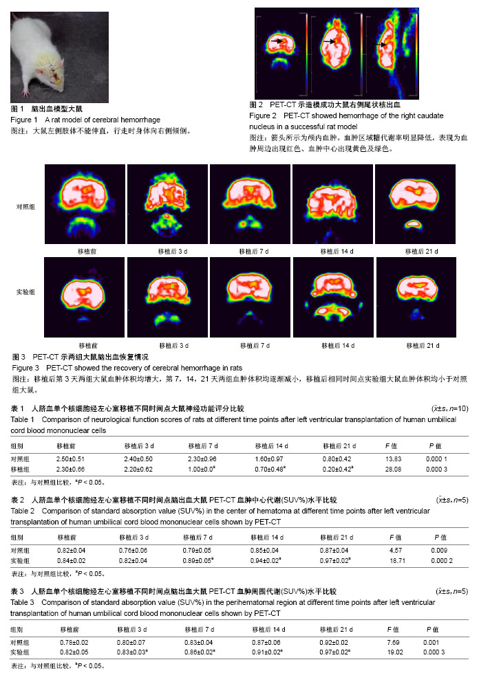
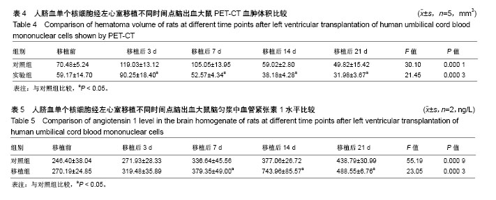
| [1] Duan X, Wen Z, Shen H, et al. Intracerebral Hemorrhage, Oxidative Stress, and Antioxidant Therapy. Oxid Med Cell Longev. 2016;2016: 1203285.[2] 魏纯纯,王培,缪朝玉. 脑出血损伤的动物模型及治疗策略的研究进展[J]. 药学实践杂志, 2016, 34(4):297-300.[3] 朱燕,邓莉,汤华军,等.电针对脑出血大鼠的实验研究[J].时珍国医国药, 2017, 28(5): 1269-1272.[4] 闫少珍,王晓莉,王海宇,等.脐血单个核细胞侧脑室移植对HIBD新生大鼠脑神经元凋亡及Bax、Bcl-2蛋白的影响[J].中国当代儿科杂志, 2016, 18(9):862-866.[5] 单泓,刘敏,韩小改,等. 脐血单个核细胞移植对大鼠脑梗死后神经功能的影响[J]. 医药论坛杂志, 2015,36(4):36-38.[6] 马泽,闫少珍,王晓莉,等.脐血单个核细胞移植联合高压氧治疗对新生大鼠缺氧缺血性脑损伤的影响[J]. 中国当代儿科杂志, 2015,17(7):736-740.[7] 张贯石,贾芙蓉,王辉,等.脐血单个核细胞治疗大脑中动脉闭塞性脑梗塞大鼠后行为学及胆碱能变化[J]. 中国免疫学杂志, 2014,30(9):1186-1188.[8] Cunha L, Horvath I, Ferreira S,et al. Preclinical imaging: an essential ally in modern biosciences. Mol Diagn Ther. 2014;18(2):153-173.[9] 杨凡慧,张春银. PET及PET/CT在脑出血中的应用[J].中国医学影像技术, 2014,30(9):1428-1431.[10] Zhou H, Tang T, Guo C,et al. Expression of Angiopoietin-1 and the receptor Tie-2 mRNA in rat brains following intracerebral hemorrhage. Acta Neurobiol Exp (Wars). 2008;68(2):147-154.[11] 实验动物管理条例[J].实用器官移植电子杂志,2016,4(2):66-67.[12] Deinsberger W, Vogel J, Kuschinsky W, et al. Experimental intracerebral hemorrhage: description of a double injection model in rats. Neurol Res. 1996;18(5):475-477.[13] Castaño J, Menendez P, Bruzos-Cidon C,et al. Fast and efficient neural conversion of human hematopoietic cells. Stem Cell Reports. 2014;3(6):1118-1131.[14] Winkler I, Kamińiska T, Bojarska-Junak A, et al. Umbilical cord blood as a source of nerve and stem cells. Ginekol Pol. 2015;86(8):603-610.[15] Liu L, Cen J, Man Y,et al. Transplantation of Human Umbilical Cord Blood Mononuclear Cells Attenuated Ischemic Injury in MCAO Rats via Inhibition of NF-κB and NLRP3 Inflammasome. Neuroscience. 2018; 369:314-324.[16] 姚星宇,杨丽敏,张国华. 人脐带血单核细胞中的神经样细胞研究[J]. 临床神经病学杂志, 2013, 26(1):41-43.[17] Boltze J, Schmidt UR, Reich DM,et al. Determination of the therapeutic time window for human umbilical cord blood mononuclear cell transplantation following experimental stroke in rats. Cell Transplant. 2012;21(6):1199-1211.[18] Song J, Li P, Chaudhary N,et al. Correlating Cerebral (18)FDG PET-CT Patterns with Histological Analysis During Early Brain Injury in a Rat Subarachnoid Hemorrhage Model. Transl Stroke Res. 2015;6(4): 290-295.[19] Zenonos G, Kim JE.18F-fluoromisonidazole PET/CT and related gauges of cerebral metabolism identify salvageable tissue in ischemic penumbra following aneurysmal subarachnoid hemorrhage. Neurosurgery.2010;66(2):N13-15.[20] 朱华,冯铭,卢珊,等.人骨髓间充质干细胞脑内移植对食蟹猴脑出血的治疗作用[J].中国实验动物学报, 2011, 19(5):361-365.[21] 林祥涛,刘树伟,刘庆伟,等.猫脑出血血肿周围组织糖代谢的(18)F-FDGPET/CT研究[J]. 中国临床解剖学杂志, 2004, 22(5):485-488.[22] Fischer M, Dietmann A, Lackner P, et al. Endovascular cooling and endothelial activation in hemorrhagic stroke patients. Neurocrit Care. 2012;17(2):224-230.[23] 胡涛.促血管生成素-1蛋白减少大鼠尿激酶溶栓后脑出血转化及机制研究[J].中国药师, 2016, 19(8):1455-1459.[24] Bihl JC, Zhang C, Zhao Y, et al. Angiotensin-(1-7) counteracts the effects of Ang II on vascular smooth muscle cells, vascular remodeling and hemorrhagic stroke: Role of the NFкB inflammatory pathway. Vascul Pharmacol. 2015;73:115-123.[25] Liao W, Xie J, Zhong J, et al. Therapeutic effect of human umbilical cord multipotent mesenchymal stromal cells in a rat model of stroke. Transplantation. 2009;87(3):350-359.[26] 姚星宇,杨丽敏,张国华.不同途径移植人脐血单核细胞对脑出血大鼠神经功能的影响[J]. 中国卒中杂志, 2012, 7(10):796-791.[27] Galieva LR, Mukhamedshina YO, Arkhipova SS, et al. Human Umbilical Cord Blood Cell Transplantation in Neuroregenerative Strategies. Front Pharmacol. 2017;8:628.[28] 唐华兵.人脐血单核细胞不同移植时间对新生大鼠缺氧缺血性脑损伤的神经保护作用研究[D].合肥:安徽医科大学, 2015.[29] 陆茵,朱霞明,李芹,等.第三方脐血输注不良反应现状及其影响因素研究[J].中华护理杂志,2017,52(5):576-580. |
.jpg)


.jpg)