中国组织工程研究 ›› 2018, Vol. 22 ›› Issue (13): 1976-1980.doi: 10.3969/j.issn.2095-4344.0491
• 骨髓干细胞 bone marrow stem cells • 上一篇 下一篇
大鼠骨髓来源内皮祖细胞体外增殖和凋亡及阿托伐他汀的干预
张日霖1,陈淑玲1,李上海2,宁奕明3,李庆军1,叶小敏1,梁伟均2
- 1湛江中心人民医院,广东省湛江市 524000;2广东医科大学附属医院,广东省湛江市 524000;3吴川市妇幼保健院,广东省吴川市 524500
Atorvastatin effects on proliferation and apoptosis of rat bone marrow-derived endothelial progenitor cells in vitro
Zhang Ri-lin1, Chen Shu-ling1, Li Shang-hai2, Ning Yi-ming3, Li Qing-jun1, Ye Xiao-min1, Liang Wei-jun2
- 1Central People’s Hospital of Zhanjiang, Zhanjiang 524000, Guangdong Province, China; 2Affiliated Hospital of Guangdong Medical University, Zhanjiang 524000, Guangdong Province, China; 3Maternal and Child Health Hospital of Wuchuan, Wuchuan 524500, Guangdong Province, China
摘要:
文章快速阅读:
.jpg)
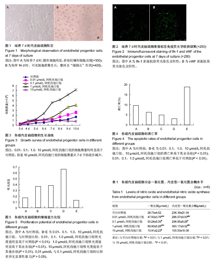
文题释义: 内皮祖细胞:是血管内皮细胞的前体,亦称成血管细胞。该细胞最早于1997年被Asahara等证明存在于循环外周血中并能分化为血管内皮细胞,在缺血刺激下可通过动员到外周血,迁移、归巢到血管新生部位,并在迁移部位增殖、分化为内皮细胞,在体内具有明显的内皮修复及促血管新生作用。多数研究认为内皮祖细胞是表达CD34、CD133、VEGFR-2、Flk -1、vWF的细胞,但具体的表达标志尚存在颇多争议,因此,其表达标志并无特异性。 内皮祖细胞移植:①内皮祖细胞的提取、鉴定、扩增上仍需一个具体和规范的方法与标准,未扩增的内皮祖细胞含量极低,而扩增后的细胞归巢能力下降,因此内皮祖细胞向心肌细胞和血管内皮细胞的分化概率也很低;②缺血缺氧状态下内皮祖细胞动员、募集和归巢受众多化学趋化因子和细胞因子的调节,它们之间的相互作用机制目前仍需进一步明确;③细胞移植时机的选择直接影响到细胞的存活。移植时间过早可能由于局部微环境恶劣致使大量移植的细胞死亡;移植时间过晚则可能导致局部损伤已不可逆;④如何保证移植后的定向分化,如分化为心肌细胞或血管内皮细胞等,且不会分化为肿瘤,这些都待进一步研究。
中图分类号:
.jpg)