中国组织工程研究 ›› 2015, Vol. 19 ›› Issue (28): 4576-4581.doi: 10.3969/j.issn.2095-4344.2015.28.027

• 干细胞培养与分化 stem cell culture and differentiation • 上一篇    下一篇

载神经生长因子纳米粒诱导神经干细胞分化为神经元及PI3K/Akt通路的影响

陈 艳1,包国庆2,刘菲菲2,张君度2,潘翠环1,龙大宏2     

  1. 1广州医科大学附属第二医院康复医学科,广东省广州市 510260;
    2广州医科大学解剖教研室,广东省广州市 510182
  • 出版日期:2015-07-02 发布日期:2015-07-02
  • 通讯作者: 龙大宏,博士,教授,广州医科大学解剖学教研室,广东省广州市 510182
  • 作者简介:陈艳,女,1976年生,湖南省沅江市人,汉族,2014年广州医科大学毕业,博士,副教授,主要从事语言、吞咽及认知障碍康复方面的研究。
  • 基金资助:

    广州医科大学青年基金项目(2011A17),项目名称:NGF与b-FGF联合诱导神经干细胞分化为胆碱能神经元

Influence of NGF-PEG-PLGA-NPs on neuronal differentiation of neural stem cells and PI3K/Akt signaling pathway   

Chen Yan1, Bao Guo-qing2, Liu Fei-fei2, Zhang Jun-du2, Pan Cui-huan1, Long Da-hong2   

  1. 1Department of Rehabilitation Medicine, the Second Affiliated Hospital of Guangzhou Medical University, Guangzhou 510260, Guangdong Province, China; 
    2Department of Anatomy, Guangzhou Medical University, Guangzhou 510182, Guangdong Province, China
  • Online:2015-07-02 Published:2015-07-02
  • Contact: Long Da-hong, M.D., Professor, Department of Anatomy, Guangzhou Medical University, Guangzhou 510182, Guangdong Province, China
  • About author:Chen Yan, M.D., Associate professor, Department of Rehabilitation Medicine, the Second Affiliated Hospital of Guangzhou Medical University, Guangzhou 510260, Guangdong Province, China
  • Supported by:

    the Youth Foundation of Guangzhou Medical University, No. 2011A17


摘要:

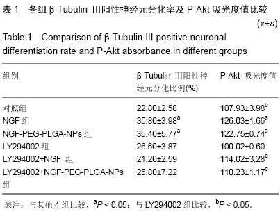
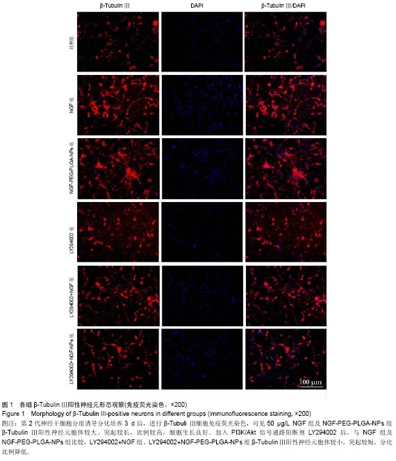
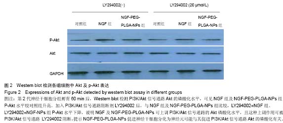
背景:课题组前期研究证实NGF-PEG-PLGA-NPs在体外有良好的缓释效能及生物活性,可以诱导PC12细胞向神经元样细胞分化。
目的:探讨NGF-PEG-PLGA-NPs诱导胎鼠大脑隔区来源神经干细胞分化为神经元的可行性及其对PI3K/Akt信号通路的影响。
方法:采用优化处方,复乳化溶剂扩散法制备NGF-PEG-PLGA-NPs。设对照组、NGF组、NGF-PEG-PLGA-NPs组、LY294002组、LY294002+NGF组、LY294002+NGF-PEG-PLGA-NPs组,诱导神经干细胞分化为神经元,细胞免疫荧光染色对神经元进行鉴定,Werstern-blotting检测PI3K/Akt信号通路Akt磷酸化水平。
结果与结论:对照组、NGF组、NGF-PEG-PLGA-NPs组、LY294002组、LY294002+NGF组、LY294002+NGF-PEG-PLGA-NPs组β-Tubulin Ⅲ阳性神经元分化率分别为(22.80±2.58)%,(35.80±3.98)%,(35.40±5.77)%,(26.60±3.87)%,(21.20±2.59)%,(25.80±7.22)%。NGF组与NGF-PEG-PLGA-NPs组神经元分化率差异无显著性意义(P > 0.05),但两组神经元分化率均高于其他各组(P < 0.05)。Western blotting检测结果显示:NGF组及NGF-PEG-PLGA-NPs组Akt磷酸化水平差异无显著性意义(P > 0.05),但均高于其他各组(P < 0.05);LY294002+NGF组及LY294002+NGF-PEG-PLGA-NPs组Akt磷酸化水平与对照组比较差异无显著性意义(P > 0.05),但均高于LY294002组(P < 0.05)。结果表明NGF-PEG-PLGA-NPs可促进神经干细胞分化为神经元,其可能机制与促进PI3K/Akt信号通路Akt的磷酸化有关。

中国组织工程研究杂志出版内容重点:干细胞;骨髓干细胞;造血干细胞;脂肪干细胞;肿瘤干细胞;胚胎干细胞;脐带脐血干细胞;干细胞诱导;干细胞分化;组织工程

关键词: 干细胞, 分化, 神经生长因子, 纳米粒, 神经干细胞, 神经元, 信号通路

Abstract:

BACKGROUND: Our previous studies confirmed that NGF-PEG-PLGA-NPs has good sustained release effect and biological activity in vitro, and can induce the differentiation of PC12 cells into neuron-like cells.
OBJECTIVE: To investigate the feasibility of neuronal differentiation of neural stem cells from septal area of fetal brain induced by NGF-PEG-PLGA-NPs and its influence on PI3K/Akt signaling pathway.
METHODS: According to optimization prescription, NGF-PEG-PLGA-NPs were prepared by multiple emulsion solvent diffusion method. Neural stem cells were induced to neuronal differentiation in six groups, including control group, NGF group, NGF-PEG-PLGA-NPs group, LY294002 group, LY294002+NGF group and LY294002+NGF-PEG-PLGA-NPs group. Neurons were identified by immunofluorescence, while phosphorylation levels of Akt in PI3K/Akt signaling pathway were detected by western blotting.
RESULTS AND CONCLUSION: The proportions of β-Tubulin III-positive neurons in control group, NGF group, NGF-PEG-PLGA-NPs group, LY294002 group, LY294002+NGF group and LY294002+NGF-PEG-PLGA-NPs group were (22.80±2.58)%, (35.80±3.98)%, (35.40±5.77)%, (26.60±3.87)%, (21.20±2.59)% and (25.80±7.22)%, respectively. There were no statistical differences in neuronal differentiation between NGF group and NGF-PEG-PLGA-NPs group
(P > 0.05), but the ratios of neural differentiation in the two groups were both higher than that in the other four groups
(P < 0.05). Western blotting results revealed that there were no statistical differences in Akt phosphorylation levels between NGF group and NGF-PEG-PLGA-NPs group (P > 0.05), but the phosphorylation levels of Akt were both higher than other four groups (P < 0.05). There were also no significant differences between LY294002+NGF and LY294002+NGF-PEG-PLGA-NPs groups and control group (P > 0.05), but the phosphorylation levels of Akt were higher than LY294002 group (P < 0.05). Results suggest that NGF-PEG-PLGA-NPs promoted neural differentiation of neural stem cells. The role might be related to increasing phosphorylation levels of Akt in PI3K/Akt signaling pathway.

中国组织工程研究杂志出版内容重点:干细胞;骨髓干细胞;造血干细胞;脂肪干细胞;肿瘤干细胞;胚胎干细胞;脐带脐血干细胞;干细胞诱导;干细胞分化;组织工程

Key words: Neural Stem Cells, Nerve Growth Factor, Nanoparticles

中图分类号: