| [1]Guo P, Huang Z, Yu P, et al. Trends in cancer mortality in China: an update. Ann Oncol. 2012; 23(10):2755-2762.
[2]Siegel R, Naishadham D, Jemal A. Cancer statistics, 2013. CA: a cancer journal for clinicians. 2013; 63(1):11-30.
[3]Nguyen LV, Vanner R, Dirks P, et al. Cancer stem cells: an evolving concept. Nat Rev Cancer. 2012; 12(2):133-143.
[4]Tomasetti C, Vogelstein B, Parmigiani G. Half or more of the somatic mutations in cancers of self-renewing tissues originate prior to tumor initiation. Proc Natl Acad Sci U S A. 2013; 110(6):1999-2004.
[5]Brenton JD, Stingl J, Stem cells: Anatomy of an ovarian cancer. Nature. 2013; 495(7440):183-184.
[6]Hammoud SS, Cairns BR, Jones DA. Epigenetic regulation of colon cancer and intestinal stem cells. Curr Opin Cell Biol. 2013;25(2):177-183.
[7]Holland JD, Klaus A, Garratt AN, et al. Wnt signaling in stem and cancer stem cells. Curr Opin Cell Biol. 2013;25(2):254- 264.
[8]Günes C, Rudolph KL. The Role of Telomeres in Stem Cells and Cancer. Cell. 2013; 152(3):390-393.
[9]Charafe-Jauffret E, Ginestier C, Bertucci F, et al. ALDH1-positive cancer stem cells predict engraftment of primary breast tumors and are governed by a common stem cell program. Cancer Res. 2013;73(24):7290-7300.
[10]Kakarala M, Brenner DE, Korkaya H, et al. Targeting breast stem cells with the cancer preventive compounds curcumin and piperine. Breast Cancer Res Treat. 2010; 122(3): 777-785.
[11]Han DW, Tapia N, Hermann A, et al. Direct reprogramming of fibroblasts into neural stem cells by defined factors. Cell Stem Cell. 2012; 10(4):465-472.
[12]Chen J, McKay RM, Parada LF. Malignant glioma: lessons from genomics, mouse models, and stem cells. Cell. 2012; 149(1):36-47.
[13]Amsterdam A, Raanan C, Schreiber L, et al. Differential localization of LGR5 and Nanog in clusters of colon cancer stem cells. Acta Histochemica. 2012.
[14]Vizio B, Mauri FA, Prati A, et al. Comparative evaluation of cancer stem cell markers in normal pancreas and pancreatic ductal adenocarcinoma. Oncol Rep. 2012; 27(1):69-76.
[15]Liang D, Shi Y. Aldehyde dehydrogenase-1 is a specific marker for stem cells in human lung adenocarcinoma. Med Oncol. 2012; 29(2):633-639.
[16]Huch M, Dorrell C, Boj SF, et al. In vitro expansion of single Lgr5+ liver stem cells induced by Wnt-driven regeneration. Nature. 2013; 494(7436):247-250.
[17]Rountree CB, Mishra L, Willenbring H. Stem cells in liver diseases and cancer: recent advances on the path to new therapies. Hepatology. 2012; 55(1):298-306.
[18]Schwartz T, Stark A, Pang J, et al. Expression of aldehyde dehydrogenase 1 as a marker of mammary stem cells in benign and malignant breast lesions of Ghanaian women. Cancer. 2013; 119(3):488-494.
[19]Smalley MJ, Kendrick H, Sheridan JM, et al. Isolation of mouse mammary epithelial subpopulations: a comparison of leading methods. J Mammary Gland Biol Neoplasia. 2012; 17(2):91-97.
[20]Guler G, Balci S, Costinean S, et al. Stem cell-related markers in primary breast cancers and associated metastatic lesions. Mod Pathol. 2012; 25(7):949-955.
[21]Sleeman KE, Kendrick H, Ashworth A, et al. CD24 staining of mouse mammary gland cells defines luminal epithelial, myoepithelial/basal and non-epithelial cells. Breast Cancer Res. 2005; 8(1):R7.
[22]Shackleton M, Vaillant F, Simpson KJ, et al. Generation of a functional mammary gland from a single stem cell. Nature. 2006; 439(7072):84-88.
[23]Liao MJ, Zhang CC, Zhou B, et al. Enrichment of a population of mammary gland cells that form mammospheres and have in vivo repopulating activity. Cancer Res. 2007; 67(17):8131- 8138.
[24]Welm BE, Tepera SB, Venezia T, et al. Sca-1(pos) cells in the mouse mammary gland represent an enriched progenitor cell population. Dev Biol. 2002; 245(1):42-56.
[25]Stingl J, Eirew P, Ricketson I, et al. Purification and unique properties of mammary epithelial stem cells. Nature. 2006; 439(7079):993-997.
[26]McQualter JL, Brouard N, Williams B, et al. Endogenous Fibroblastic Progenitor Cells in the Adult Mouse Lung Are Highly Enriched in the Sca‐1 Positive Cell Fraction. Stem Cells. 2009; 27(3):623-633.
[27]DeOme K, Faulkin L, Bern HA, et al. Development of mammary tumors from hyperplastic alveolar nodules transplanted into gland-free mammary fat pads of female C3H mice. Cancer Res. 1959; 19(5):515.
[28]Regan J, Smalley M. Prospective isolation and functional analysis of stem and differentiated cells from the mouse mammary gland. Stem Cell Rev. 2007; 3(2):124-136.
[29]Staszkiewicz J, Gimble JM, Manuel JA, et al. IFATS Collection: Stem Cell Antigen-1-Positive Ear Mesenchymal Stem Cells Display Enhanced Adipogenic Potential. Stem Cells. 2008; 26(10):2666-2673.
[30]Kang SG, Shinojima N, Hossain A, et al. Isolation and perivascular localization of mesenchymal stem cells from mouse brain. Neurosurgery. 2010; 67(3):711.
[31]Naftali‐Shani N, Itzhaki‐Alfia A, Landa‐Rouben N, et al. The Origin of Human Mesenchymal Stromal Cells Dictates Their Reparative Properties. J Am Heart Assoc. 2013; 2(5): e000253.
[32]Tan KY, Eminli S, Hettmer S, et al. Efficient generation of iPS cells from skeletal muscle stem cells. PloS one. 2011; 6(10): e26406.
[33]Suzuki K, Sun R, Origuchi M, et al. Mesenchymal stromal cells promote tumor growth through the enhancement of neovascularization. Molecular Med. 2011; 17(7-8):579.
[34]Siegel G, Krause P, Wöhrle S, et al. Bone marrow-derived human mesenchymal stem cells express cardiomyogenic proteins but do not exhibit functional cardiomyogenic differentiation potential. Stem Cells Dev. 2012; 21(13): 2457-2470.
[35]Sakakura T, Suzuki Y, Shiurba R. Mammary stroma in development and carcinogenesis. J Mammary Gland Biol Neoplasia. 2013:1-9.
[36]dos Santos CO, Rebbeck C, Rozhkova E, et al. Molecular hierarchy of mammary differentiation yields refined markers of mammary stem cells. Proc Natl Acad Sci. 2013; 110(18): 7123-7130.
[37]Roy S, Gascard P, Dumont N, et al. Rare somatic cells from human breast tissue exhibit extensive lineage plasticity. Proc Natl Acad Sci. 2013; 110(12):4598-4603.
[38]Fan Y, Menon RK, Cohen P, et al. Liver-specific deletion of the growth hormone receptor reveals essential role of growth hormone signaling in hepatic lipid metabolism. J Biol Chem. 2009; 284(30):19937-19944.
[39]Horigan K, Trott J, Barndollar A, et al. Hormone interactions confer specific proliferative and histomorphogenic responses in the porcine mammary gland. Domest Anim Endocrinol. 2009; 37(2):124-138.
[40]Cheng G, Weihua Z, Warner M, et al. Estrogen receptors ER alpha and ER beta in proliferation in the rodent mammary gland. Proc Natl Acad Sci U S A. 2004; 101(11):3739-3746.
[41]Morani A, Warner M, Gustafsson JA. Biological functions and clinical implications of oestrogen receptors alfa and beta in epithelial tissues. J Intern Med. 2008; 264(2):128-142.
[42]Clarke RB, Spence K, Anderson E, et al. A putative human breast stem cell population is enriched for steroid receptor-positive cells. Dev Biol. 2005; 277(2):443-456.
[43]Booth BW, Smith GH. Estrogen receptor-alpha and progesterone receptor are expressed in label-retaining mammary epithelial cells that divide asymmetrically and retain their template DNA strands. Breast Cancer Res. 2006; 8(4): R49.
[44]Booth BW, Boulanger CA, Smith GH. Selective segregation of DNA strands persists in long-label-retaining mammary cells during pregnancy. Breast Cancer Res. 2008; 10(5):R90.
[45]Wang S, Qu X, Zhao RC. Clinical applications of mesenchymal stem cells. J Hematol Oncol. 2012; 5(1):19. |
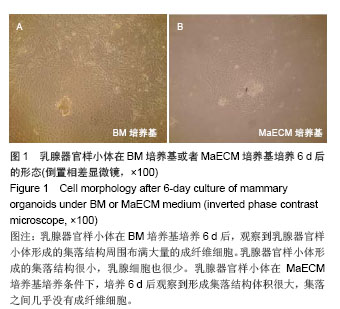
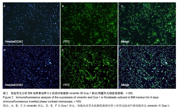
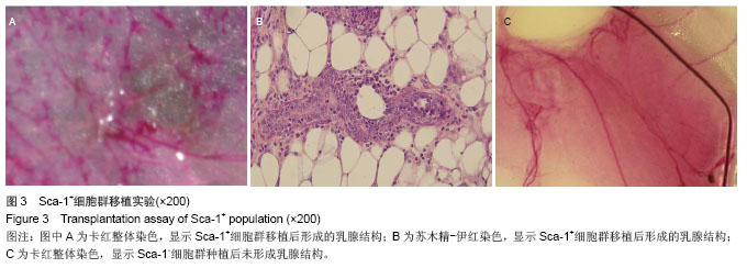
.jpg)