[1] Chen G, Tang Q, Yu S, et al. The biological function of Bmal1 in skeleton development and disorders. Life Sci. 2020;3:117636.
[2] Shimizu T, Watanabe K, Anayama N, et al. Effect of lipopolysaccharide on circadian Clock genes Per2 and Bmal1 in mouse ovary. J Physiol Sci. 2017;67(5):623-628.
[3] Yoshida K, Nakai A, Kaneshiro K, et al.TNF-a induces expression of the circadian Clock gene Bmal1 via dual calcium-dependent pathways in rheumatoid synovial cells. Biochem Biophys Res Commun. 2018;495(2):1675-1680.
[4] Mayeuf-Louchart A, Staels B, Duez H. Skeletal muscle functions around the Clock. Diabetes Obes Metab. 2015;17 Suppl 1:39-46.
[5] Gibbs JE, Ray DW. The role of the circadian Clock in rheumatoid arthritis. Arthritis Res Ther. 2013;15(1):205.
[6] Wang Y, Pati P, Xu Y, et al. Endotoxin Disrupts Circadian Rhythms in Macrophages via Reactive Oxygen Species. PLoS One. 2016;11(5): e0155075.
[7] Meng ZX, Gong J, Chen Z, et al. Glucose Sensing by Skeletal Myocytes Couples Nutrient Signaling to Systemic Homeostasis.Mol Cell. 2017;66(3):332-344.
[8] Hodge BA, Zhang X, Gutierrez-Monreal MA, et al. MYOD1 functions as a Clock amplifier as well as a critical co-factor for downstream circadian gene expression in muscle. Elife. 2019;8:e43017.
[9] Wang C, Liu W, Nie Y, et al. Loss of MyoD promotes fate transdifferentiation of myoblasts into brown adipocytes. EBioMedicine. 2017;16:212-223.
[10] Yoo YM, Jung EM, Jeung EB. Rapamycin-induced autophagy decreases Myf5 and MyoD proteins in C2C12 myoblast cells. Toxicol In Vitro. 2019;58:132-141.
[11] Tu C, Bu Y, Vujcic M, et al. Ion Current-based Proteomic Profiling in Understanding the Inhibitory Effect of Tumor Necrosis Factor Alpha on Myogenic Differentiation. J Proteome Res. 2016;15(9):3147-3157.
[12] Chatterjee S, Ma K. Circadian Clock regulation of skeletal muscle growth and repair. F1000Res. 2016;5:1549.
[13] Riley LA, Esser KA. The Role of the Molecular Clock in Skeletal Muscle and What It Is Teaching Us About Muscle-Bone Crosstalk. Curr Osteoporos Rep. 2017;15(3):222-230.
[14] Aoyama S, Shibata S. The Role of Circadian Rhythms in Muscular and Osseous Physiology and Their Regulation by Nutrition and Exercise.Front Neurosci. 2017;11:63.
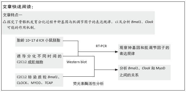
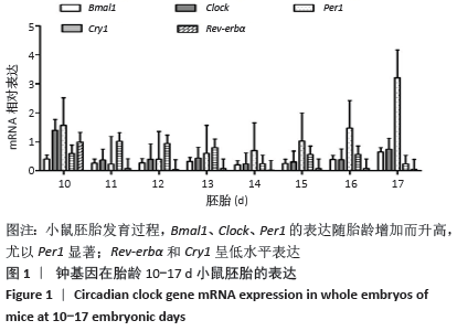
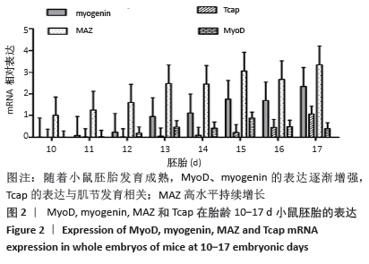
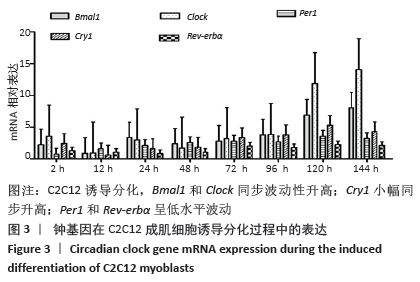
[15] 闫银弟,罗旭光,杨艳萍,等.钟基因在不同胎龄小鼠胚胎和孕鼠的表达规律[J].中国组织工程研究,2019,23(1):96-102.
[16] Chatterjee S, Yin H, Nam D, et al. Brain and muscle Arnt-like 1 promotes skeletal muscle regeneration through satellite cell expansion.Exp Cell Res. 2015;331(1):200-210.
[17] Chatterjee S, Yin H, Li W, et al. The Nuclear Receptor and Clock Repressor Rev-erbα Suppresses Myogenesis. Sci Rep. 2019;9(1): 4585.
[18] Samant SA, Kanwal A, Pillai VB, et al. The histone deacetylase SIRT6 blocks myostatin expression and development of muscle atrophy. Sci Rep. 2017;7(1):11877.
[19] 闫银弟,罗旭光,杨艳萍,等.骨骼肌昼夜节律分子钟机制的研究进展[J].实用医学杂志,2018,34(7):1213-1215.
[20] Romagnoli C, Zonefrati R, Sharma P, et al. Characterization of Skeletal Muscle Endocrine Control in an In Vitro Model of Myogenesis.Calcif Tissue Int. 2020;107(1):18-30.
[21] ROBINSON I, REDDY AB. Molecular mechanisms of the circadian Clock work in mammals. FEBS Lett. 2014;588(15):2477-2483.
[22] Armand AS, Bourajjaj M, Martinez-Martinez S, et al. CooPerative synergy between NFAT and MyoD regulates myogenin expression and myogenesis. J Biol Chem. 2008;283(43):29004-29010.
[23] de la Serna IL, Ohkawa Y, Berkes CA, et al. MyoD Targets Chromatin Remodeling Complexes to the Myogenin Locus Prior to Forming a Stable DNA-Bound Complex. Mol Cell Biol. 2005;25(10): 3997-4009.
[24] Ganassi M, Badodi S, Ortuste Quiroga HP, et al. Myogenin promotes myocyte fusion to balance fibre number and size. Nat Commun. 2018;9(1):4232.
[25] Higashioka K, Koizumi N, Sakurai H, et al. Myogenic differentiation from MYOGENIN-mutated human iPS cells by CRISPR/Cas9.Stem Cells Int. 2017;2017:9210494.
[26] Himeda CL, Ranish JA, Hauschka SD. Quantitative proteomicidentification of MAZ as a transcriptional regulator of muscle-specific genes in skeletal and cardiac myocytes. Mol Cell Biol. 2008;28(20):6521-6535.
[27] Shavlakadze T, Anwari T, Soffe Z, et al. Impact of fasting on the rhythmic expression of myogenic and metabolic factors in skeletal muscle of adult mice. Am J Physiol Cell Physiol. 2013;305(1):C26-C35.
[28] Vogel C, Marcotte EM. Insights into the regulation of protein abundance from proteomic and transcriptomic analyses.Nat Rev Genet. 2012;13(4):227-232.
[29] Sabari BR, Dall’Agnese A, Boija A, et al. Coactivator condensation at suPer-enhancers links phase separation and gene control. Science. 2018;361(6400).pii: eaar3958.
[30] Boeynaems S, Alberti S, Fawzi NL, et al. Protein phase separation: A new phase in cell biology. Trends Cell Biol. 2018;28(6):420-435.
[31] Leong I. Muscle circadian Clock regulates lipid storage.Nat Rev Endocrinol. 2018;14(10):563.
[32] Vitale JA, Bonato M, La Torre A, et al. The Role of the Molecular Clock in Promoting Skeletal Muscle Growth and Protecting against Sarcopenia. Int J Mol Sci. 2019;20(17).pii: E4318.
[33] Qiao M, Huang J, Wu H, et al. Molecular characterization, transcriptional regulation and association analysis with carcass traits of porcine TCAP gene. Gene. 2014;538(2):273-279.
[34] Zhang X, Patel SP, McCarthy JJ, et al. A non-canonical E-box within the MyoD core enhancer is necessary for circadian expression in skeletal muscle. Nucleic Acids Res. 2012;40(8):3419-3430.
|