[1] 杨丰建,俞永林,夏军,等.基质金属蛋白酶-1/13与信号通路激酶ERKl/2在兔骨关节炎软骨中的表达[J].老年医学与保健,2007, 13(6):338-342.
[2] LORIES RJ, MONTEAGUDO S. Review Article:Is Wnt Signaling an Attractive Target for the Treatment of Osteoarthritis?Rheumatol Ther. 2020;7(2):259-270.
[3] HUNTER DJ, BIERMA-ZEINSTRA S. Osteoarthritis. Lancet. 2019;393 (10182):1745-1759.
[4] XI Y, HUANG X, TAN G, et al. Protective effects of Erdosteine on interleukin-1β-stimulated inflammation via inhibiting the activation of MAPK,NF-κB,and Wnt/β-catenin signaling pathways in rat osteoarthritis. Eur J Pharmacol. 2020;873:172925.
[5] LI H, PENG Y, WANG XI, et al. Astragaloside inhibits IL-1β-induced inflammatory response in human osteoarthritis chondrocytes and ameliorates the progression of osteoarthritis in mice.Immunopharmacol Immunotoxicol. 2019;41:497-503.
[6] SCURUCHI M, D’ASCOLA A, AVENOSO A, et al. Serglycin as part of IL-1beta induced inflammation in human chondrocytes. Arch Biochem Biophys. 2019;669:80-86.
[7] KAPOOR M, MARTEL-PELLETIER J, LAJEUNESSE D, et al. Role of proinflammatory cytokines in the pathophysiology of osteoarthritis. Nat Rev Rheumatol. 2011;7:33–42.
[8] 周炎,邓明,贺斌,等.大鼠诱发性骨关节炎模型构建研究进展[J].国际骨科学杂志,2016,37(5):304-310.
[9] ONISHI RM, GAFFEN SL. Interleukin-17 and its target genes: mechanisms of interleukin-17 function in disease.Immunology. 2010; 129:311-321.
[10] LIU Y, PENG H, MENG Z, et al. Correlation of IL-17 Level in Synovia and Severity of Knee Osteoarthritis.Med Sci Monit. 2015;21:1732-1736.
[11] LEE J, LEE SY, KANG CM, et al. Interleukin-17 Enhances Germinal Center Formation and Immunoglobulin G1 Production in Mice.Rheum Dis. 2017;24:271-278.
[12] ZHANG J, LI Q, CHANG S. The effects of particle density in moxa smoke on the ultrastructure of knee cartilage and expressions of TNF-α,IL-1b,BAX,and Bcl-2 mRNA in a rat model for osteoarthritis.J Cell Biochem. 2019;120(4):6589-6595.
[13] 陈波,谢西梅,李佳霖.艾灸治疗对兔 KOA 软骨损伤及TNF-α、TGF-β1 和 IGF-1表达调节作用的实验研究[J].江苏中医药,2009, 41(6):69-71.
[14] LIAO C, WANG S, ZHU S, et al. Advanced oxidation protein products increase TNF-α and IL-1β expression in chondrocytes via NADPH oxidase 4 and accelerate cartilage degeneration in osteoarthritis progression. Redox Biol. 2020;28:101306.
[15] MEDVEDEVA EV, GREBENIK EA, GORNOSTAEVA SN, et al. Repair of Damaged Articular Cartilage:Current Approaches and Future Directions. Int J Mol Sci. 2018;19(8):2366.
[16] 郭伟雄,魏波.炎症细胞因子及通路在骨关节炎中的研究进展[J].国际检验医学杂志,2015,36(15):2240-2241.
[17] 李灿锋,陈卓,曾羿,等.骨形态发生蛋白信号通路在骨关节炎发病机制中的作用研究[J].中华关节外科杂志(电子版),2017,11(5): 500-505.
[18] CHIEN SY, TSAI CH, LIU SC, et al. Noggin Inhibits IL-1β and BMP-2 Expression,and Attenuates Cartilage Degeneration and Subchondral Bone Destruction in Experimental Osteoarthritis. Cells. 2020;9(4):E927.
[19] THIELEN NGM, VAN DER KRAAN PM, VAN CAAM APM. TGFβ/BMP Signaling Pathway in Cartilage Homeostasis. Cells. 2019;8(9):969.
[20] CHOI B, KIM S, FAN J, et al. Covalently conjugated transforming growth factor-β1 in modular chitosan hydrogels for the effective treatment of articular cartilage defects. Biomater Sci. 2015;3(5):742-752.
[21] RUIZ M, MAUMUS M, FONTENEAU G, et al. TGFβi is involved in the chondrogenic differentiation of mesenchymal stem cells and is dysregulated in osteoarthritis. Osteoarthr Cartil. 2019;27:493-503.
[22] 樊志强,庞炜,杨连甲,等.白介素1受体拮抗剂及转化生长因子β1对兔膝关节骨性关节炎(OA)的治疗研究[J].现代生物医学进展, 2011,11(13):2447-2450.
[23] WIEGERTJES R, VAN CAAM A, VAN BEUNINGEN H, et al. TGF-β dampens IL-6 signaling in articular chondrocytes by decreasing IL-6 receptor expression. Osteoarthr Cartil. 2019;27:1197-1207.
[24] VAN DER KRAAN PM. The changing role of TGFbeta in healthy,ageing and osteoarthritic joints.Nat Rev Rheumatol. 2017;13:155-163.
[25] CUNHA SI, PIETRAS K. ALK1 as an emerging target for antiangiogenic therapy of cancer. Blood. 2011;117:6999-7006.
[26] TAN Y, LU K, LI J, et al. Prenatal caffeine exprosure increases adult female offspring rat’s susceptibility to osteoarthritis via low-functional programming of cartilage IGF-1 with histone acetylation. Toxicol Lett. 2018;295:229-236.
[27] PASOLD J, ZANDER K, HESKAMP B, et al. Positive impact of IGF-1-coupled nanoparticles on the differentiation potential of human chondrocytes cultured on collagen scaffolds. Int J Nanomedicine. 2015;10:1131-1143.
[28] UCHIMURA T, FOOTE AT, SMITH EL, et al. Insulin-Like Growth Factor II (IGF-II) Inhibits IL-1β-Induced Cartilage Matrix Loss and Promotes Cartilage Integrity in Experimental Osteoarthritis. J Cell Biochem. 2015; 116(12):2858-2869.
[29] SCHWARZ S, MROSEWSKI I, SILAWAL S, et al. The interrelation of osteoarthritis and diabetes mellitus:considering the potential role of interleukin-10 and in vitro models for further analysis. Inflamm Res. 2018;67(4):285‐300.
[30] BEHRENDT P, PREUSSE-PRANGE A, KLÜTER T, et al. IL-10 reduces apoptosis and extracellular matrix degradation after injurious compression of mature articular cartilage. Osteoarthritis Cartilage. 2016;24(11):1981-1988.
[31] VAN MEEGEREN ME, ROOSENDAAL G, COELEVELD K, et al. A single intra-articular injection with IL-4 plus IL-10 ameliorates blood-induced cartilage degeneration in haemophilic mice.Br J Haematol. 2013;160(4):515-520.
[32] SONG SY, HONG J, GO S, et al. Interleukin-4 Gene Transfection and Spheroid Formation Potentiate Therapeutic Efficacy of Mesenchymal Stem Cells for Osteoarthritis. Adv Healthc Mater. 2020;9(5):e1901612.
[33] WANG J, CHEN H, CAO P, et al. Inflammatory cytokines induce caveolin-1/β-catenin signalling in rat nucleus pulposus cell apoptosis through the p38 MAPK pathway. Cell Prolif. 2016;49(3):362-372.
[34] Yao J, Weng Y, Yan S, et al. NOV inhibits proliferation while promoting apoptosis and migration in osteosarcoma cell lines through p38/MAPK and JNK/MAPK pathways. Oncology Reports. 2015;34(4): 2011-2021.
[35] SUN HY, HU KZ, YIN ZS. Inhibition of the p38-MAPK signaling pathway suppresses the apoptosis and expression of proinflammatory cytokines in human osteoarthritis chondrocytes. Cytokine. 2017;90:135-143.
[36] PRASADAM I, MAO X, SHI W, et al. Combination of MEK ERK inhibitor and hyaluronic acid has a synergistic effect on antihypertrophic and pro chondrogenic activities in osteoarthritis treatment. J Mol Med(Berl). 2013;91(3):369-380.
[37] GARCÍA-VELÁZQUEZ L, ARIAS C. The emerging role of Wnt signaling dysregulation in the understanding and modification of age-associated diseases. Ageing Res Rev. 2017;37:135-145.
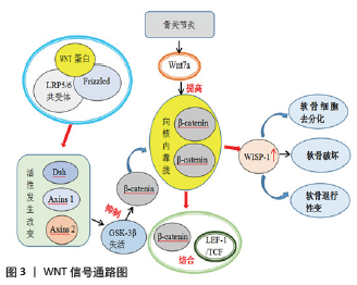
[38] ZHOU Y, WANG T, HAMILTON JL, et al. Wnt/β-catenin Signaling in Osteoarthritis and in Other Forms of Arthritis. Curr Rheumatol Rep. 2017;19(9):53.
[39] STAMPELLA A, MONTEAGUDO S, LORIES R. Wnt signaling as target for the treatment of osteoarthritis. Best Pract Res Clin Rheumatol. 2017;31(5):721-729.
[40] SASSI L, LAADHAR M, ALLOUCHE B, et al. The roles of canonical and non-canonical Wnt signaling in human de-differentiated articular chondrocytes. Biotech Histochem. 2014;89(1):53-65.
[41] 唐萌芽,倪慧英,张学民,等.补肾活血中药对膝骨关节炎患者经典 Wnt /β-catenin 通路调控作用的临床研究[J].中医正骨,2014, 26(8):12-14+17.
[42] BLOM AB, BROCKBANK SM, VAN LENT PL, et al. Involvement of the Wnt signaling pathway in experimental and human osteoarthritis: prominent role of Wnt-induced signaling protein 1. Arthritis Rheum. 2009;60( 2):501-512.
[43] LIETMAN C, WU B, LECHNER S, et al. Inhibition of Wnt/β-catenin signaling ameliorates osteoarthritis in a murine model of experimental osteoarthritis. JCI Insight. 2018;3(3):e96308.
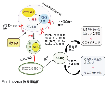
[44] 张清,向明,陈杭,等.Notch 信号通路在骨关节炎软骨细胞中的表达及意义[J].局解手术学杂志,2018,27(4):240-245.
[45] KOVALL RA, GEBELEIN B, SPRINZAK D, et al. The canonical notch signaling pathway:structural and biochemical insights into shape, sugar, and force. Developmental Cell. 2017;41(3):228.
[46] LIU Z, REN Y, MIRANDO AJ, et al. Notch signaling in postnatal joint chondrocytes,but not subchondral osteoblasts,is required for articular cartilage and joint maintenance. Osteoarthritis Cartilage. 2016;24(4): 740-751.
[47] ZHENG Y, LIU C, NI L, et al. Cell type-specific effects of Notch signaling activation on intervertebral discs:Implications for intervertebral disc degeneration. J Cell Physiol. 2018;233(7):5431-5440.
[48] Mahjoub M, Sassi N, Driss M, et al. Expression patterns of Notch receptors and their ligands in osteoarthritic and healthy human knee cartilage. Tissue and Cell. 2012;44(3):182-194.
[49] 吴绍军,刘俊才,左银龙,等.Notch信号通路在膝骨关节炎软骨细胞凋亡中的作用研究[J].华西医学,2018,33( 9):1162 -1167.
[50] Liu Z, Chen J, Mirando AJ, et al. A dual role for NOTCH signaling in joint cartilage maintenance and osteoarthritis. Sci Signal. 2015; 8(386):71-76.
[51] Rigoglou S, Papavassiliou AG. The NF-κB signalling pathway in osteoarthritis. Int J Biochem Cell Biol. 2013;45(11):2580-2584.
[52] Ji B, Guo W, Ma H, et al. Isoliquiritigenin suppresses IL-1β induced apoptosis and inflammation in chondrocyte-like ATDC5 cells by inhibiting NF-κB and exerts chondroprotective effects on a mouse model of anterior cruciate ligament transection. Int J Mol Med. 2017; 40(6):1709-1718.
[53] Dai T, Shi K, Chen G, et al. Malvidin attenuates pain and inflammation in rats with osteoarthritis by suppressing NF-κB signaling pathway. Inflamm Res. 2017;66(12):1075-1084.
[54] Ma D, Zhao Y, She J, et al. NLRX1 alleviates lipopolysaccharide-induced apoptosis and inflammation in chondrocytes by suppressing the activation of NF-κB signaling. International Immunopharmacology. 2019;71:7-13.
[55] Pan T, Chen R, Wu D, et al. Alpha-Mangostin suppresses interleukin-1β-induced apoptosis in rat chondrocytes by inhibiting the NF-κB signaling pathway and delays the progression of osteoarthritis in a rat model. International Immunopharmacology. 2017;52:156-162.
|
 文题释义:
文题释义: