[1] 张世民, 胡孙君, 张立智,等.老年髋部转子间骨折[M]. 北京: 科学出版社.2019.
[2] LEUNG F, BLAUTH M, BAVONRATANAVECH S. Surgery for fragility hip fracture-streamlining the process. Osteoporos Int. 2010;21(Suppl 4): S519-521.
[3] 曹军, 胡攀勇, 蔡保塔, 等. 三种植入物内固定治疗老年股骨转子间骨折的比较[J].中国组织工程研究,2018,22(11):1683-1688.
[4] NING GZ, LI YL, WU Q, et al. Cemented versus uncemented hemiarthroplasty for displaced femoral neck fractures: an updated meta-analysis. Eur J Orthop Surg Traumatol. 2014;24(1):7-14.
[5] HIRAGAMI K, ISHII J. Embedding the lateral end of the lag screw within the lateral wall in the repair of reverse obliquity intertrochanteric femur fracture. J Int Med Res. 2018;46(3): 1103-1108.
[6] 蔡振存,朴成哲,陈拥,等. PFNA固定治疗老年人内侧壁不完整的股骨粗隆间骨折[J]. 沈阳医学院学报,2016,18(4):253-255.
[7] HAMMER A.The calcar femorale: A new perspective. J Orthop Surg (Hong Kong) 2019;27(2):2309499019848778.
[8] ARIRACHAKARAN A, AMPHANSAP T, THANINDRATARN P, et al. Comparative outcome of PFNA, Gamma nails, PCCP, Medoff plate, LISS and dynamic hip screws for fixation in elderly trochanteric fractures: a systematic review and network meta-analysis of randomized controlled trials. Eur J Orthop Surg Traumatol. 2017;27(7):937-952.
[9] AGRAWAL P, GABA S, DAS S, et al. Dynamic hip screw versus proximal femur locking compression plate in intertrochanteric femur fractures (AO 31A1 and 31A2): A prospective randomized study. J Nat Sci Biol Med. 2017;8(1):87-93.
[10] 徐高翔,李建涛,张浩,等.中老年股骨转子间骨折患者股骨近端解剖参数的性别和年龄差异研究[J].中华创伤骨科杂志,2020, 22(3):224-231.
[11] ISIDA R, BARIATINSKY V, KERN G, et al. Prospective study of the reproducibility of X-rays and CT scans for assessing trochanteric fracture comminution in the elderly: a series of 110 cases. Eur J Orthop Surg Traumatol. 2015;25(7):1165-1170.
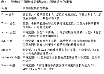
[12] 王人楷,章浩,李迪,等. 股骨粗隆间骨折临床分型研究进展[J].中国矫形外科杂志,2018,26(20):1882-1887.
[13] JENSEN JS. Classification of trochanteric fractures. Acta Orthop Scand.1980;51(5):803-810.
[14] Kyle RF, Gustilo RB, Premer RF. Analysis of six hundred and twenty-two intertrochanteric hip fractures. J Bone Joint Surg Am. 1979; 61(2):216-221.
[15] NEWMAN RJ. The comprehensive classification of fractures of long bone. Current Orthopaeclics. 1991;5(4):287.
[16] MATRE K, VINJE T, HAVELIN LI, et al. TRIGEN INTERTAN intramedullary nail versus sliding hip screw: a prospective, randomized multicenter study on pain, function, and complications in 684 patients with an intertrochanteric or subtrochanteric fracture and one year of follow-up.J Bone Joint Surg Am. 2013;95(3):200-208.
[17] SHODA E, KITADA S, SASAKI Y, et al. Proposal of new classification of femoral trochanteric fracture by three-dimensional computed tomography and relationship to usual plain X-ray classification. J Orthop Surg (Hong Kong). 2017;25(1):2309499017692700.
[18] 王郑浩,李开南,兰海.基于三维CT的股骨转子间骨折后内侧壁骨折地图的研究[J].中华创伤骨科杂志,2019,21(9):745-751.
[19] KELLAM JAMES F, MEINBERG ERIC G, JULIE A, et al. Introduction: Fracture and Dislocation Classification Compendium-2018: International Comprehensive Classification of Fractures and Dislocations Committee. J Orthop Trauma. 2018;32( Suppl 1):S1–S10.
[20] 李双, 张世民,张立智,等.不同组合前内侧皮质支撑复位对股骨转子间骨折髓内钉术后稳定性影响的生物力学研究[J]. 中华创伤骨科杂志,2019;21(1):57-64.
[21] Chang SM, Zhang YQ, Du SC, et al. Anteromedial cortical support reduction in unstable pertrochanteric fractures: a comparison of intra-operative fluoroscopy and post-operative three dimensional computerised tomography reconstruction. Int Orthop. 2018;42(1):183-189.
[22] Nie B, Chen X, Li J, et al. The medial femoral wall can play a more important role in unstable intertrochanteric fractures compared with lateral femoral wall: a biomechanical study. J Orthop Surg Res. 2017;12(1):197.
[23] 闵重函, 周瑛, 张洪美, 等. 老年国人股骨近端生物力学特点与股骨粗隆间骨折内固定选择的试验研究[J]. 中国骨与关节损伤杂志,2018,33(8):789-793.
[24] SEKER A, BAYSAL G, BILSEL N, et al. Should early weightbearing be allowed after intramedullary fixation of trochanteric femur fractures? A finite element study. J Orthop Sci. 2020;25(1):132-138.
[25] TAN BY, LAU AC, KWEK EB. Morphology and fixation pitfalls of a highly unstable intertrochanteric fracture variant. J Orthop Surg (Hong Kong). 2015;23(2):142-145.
[26] ZHANG Q, CHEN W, LIU HJ, et al. The role of the calcar femorale in stress distribution in the proximal femur. Orthop Surg. 2009;1(4):311-316.
[27] DOMINGO LJ, CECILIA D, HERRERA A, et al. Trochanteric fractures treated with a proximal femoral nail. Int Orthop. 2001;25(5): 298-301.
[28] 曹培锋,洪勇平,王以近,等.股骨小转子缺损及复位固定的生物力学比较[J].中国矫形外科杂志,2009,17(22):1722-1724.
[29] HARRINGTON KD, JOHNSTON JO. The management of comminuted unstable intertrochanteric fractures. J Bone Joint Surg Am. 1973;55(7): 1367-1376.
[30] KIM JW, LEE JI, PARK KC. Pseudoaneurysm of the deep femoral artery caused by a guide wire following femur intertrochanteric fracture with a hip nail: A case report. Acta Orthop Traumatol Turc. 2017;51(3):266-269.
[31] EHRNTHALLER C, OLIVIER AC, GEBHARD F, et al. The role of lesser trochanter fragment in unstable pertrochanteric A2 proximal femur fractures - is refixation of the lesser trochanter worth the effort. Clin Biomech (Bristol, Avon). 2017;42:31-37.
[32] 熊蠡茗, 胡益强, 邵增务, 等. 股骨近端锁定板固定治疗老年不稳定型股骨转子间骨折[J].中华创伤骨科杂志,2017,19(2):115-120.
[33] 左进步,余磊, 梁宏伟,等.钉板固定、髓内固定及人工股骨头置换修复高龄股骨转子间骨折:选择与比较[J]. 中国组织工程研究, 2015,19(17):2711-2718.
[34] JACOB J, DESAI A, TROMPETER A. Decision Making in the Management of Extracapsular Fractures of the Proximal Femur - is the Dynamic Hip Screw the Prevailing Gold Standard. Open Orthop J. 2017;11:1213-1217.
[35] MATAR HE, CHANDRAN P. Outcomes of internal fixation of intracapsular hip fractures using dynamic locking plate system (Targon® FN). J Orthop. 2018;15(3):829-831.
[36] 郭晓泽,章莹, 肖进. 动力髋螺钉固定股骨转子间骨折的有限元分析[J]. 临床骨科杂志, 2012,15(4):458-461.
[37] SIWACH RC, ROHILLA R, SINGH R, et al. Radiological and functional outcome in unstable, osteoporotic trochanteric fractures stabilized with dynamic helical hip system. Strategies Trauma Limb Reconstr. 2013;8(2):117-122.
[38] 张一鹏, 刘伟新, 梁献会. DHS治疗股骨转子间骨折失败原因分析[J]. 实用骨科杂志,2014,20(2):165-168.
[39] 陆勇. 动力髋螺钉治疗股骨粗隆间骨折失败原因分析[J]. 骨与关节损伤杂志,2003,18(11):730-732.
[40] YOO JI, HA YC, LIM JY, et al. Early Rehabilitation in Elderly after Arthroplasty versus Internal Fixation for Unstable Intertrochanteric Fractures of Femur: Systematic Review and Meta-Analysis.J Korean Med Sci. 2017;32(5):858-867.
[41] 徐锋,吕建元,洪嵘,等.植骨在DHS固定治疗股骨粗隆间骨折中的作用[J]. 实用骨科杂志, 2008,14(12):722-725.
[42] 罗小荣, 梁路石. 应用动力髋螺钉系统结合小粗隆复位固定治疗股骨转子间骨折[J]. 中国骨与关节损伤杂志,2006,21(8):659-660.
[43] 张松,王晓,尹桂荣,等.小粗隆拉力螺钉固定、植骨结合DHS治疗不稳定型股骨粗隆间骨折[J].骨与关节损伤杂志,2004,19(9):618-619.
[44] 陈燕才. 动力髋螺钉联合钢丝对股骨粗隆间骨折合并内侧壁骨折疗效分析[J].现代诊断与治疗,2017,28(4):607-609.
[45] Ceynowa M, Zerdzicki K, Klosowski P, et al. The early failure of the gamma nail and the dynamic hip screw in femurs with a wide medullary canal. A biomechanical study of intertrochanteric fractures. Clin Biomech (Bristol, Avon). 2020;71:201-207.
[46] 葛旻, 程飚, 陈富强, 等. 骨水泥强化动力髋螺钉内固定治疗老年股骨粗隆间骨折的疗效观察[J].中国骨与关节损伤杂志, 2017, 32(10):1066-1067.
[47] MÜLLER F, DOBLINGER M, KOTTMANN T, et al. PFNA and DHS for AO/OTA 31-A2 fractures: radiographic measurements, morbidity and mortality. Eur J Trauma Emerg Surg. 2019 Oct 31. doi: 10.1007/s00068-019-01251-w.
[48] WIRTZ C, ABBASSI F, EVANGELOPOULOS DS, et al. High failure rate of trochanteric fracture osteosynthesis with proximal femoral locking compression plate. Injury. 2013;44(6):751-756.
[49] 张施展, 张卫国, 徐钧, 等. 结构性植骨联合锁定钢板治疗股骨距粉碎性股骨转子间骨折的临床研究[J]. 重庆医学,2017,46(14):1919-1921.
[50] 房冰,李晓波,王硕磊.不同固定方式修复老年股骨转子间骨折的临床对比研究[J].实用老年医学 2017,31(6):551-554.
[51] 张斌,常军,杨志刚,等. 内侧壁缺损面积对股骨转子间骨折经皮加压钢板固定术后断端稳定性影响的实验研究[J].中华创伤骨科杂志,2016,18(1):61-65.
[52] LONG H, LIN Z, LU B, et al. Percutaneous compression plate versus dynamic hip screw for treatment of intertrochanteric hip fractures: A overview of systematic reviews and update meta-analysis of randomized controlled trials. Int J Surg. 2016;33 Pt A:1-7.
[53] 白浪, 侯毅龙, 张晟, 等. 三种内固定方式治疗内侧壁缺损的不稳定型股骨转子间骨折的疗效比较[J]. 中华创伤骨科杂志,2018, 20(5):412-418.
[54] SANDERS D, BRYANT D, TIESZER C, et al. A Multicenter Randomized Control Trial Comparing a Novel Intramedullary Device (InterTAN) Versus Conventional Treatment (Sliding Hip Screw) of Geriatric Hip Fractures. J Orthop Trauma.2017;31(1):1-8.
[55] 史彦海. 伴内侧壁和外侧壁缺损老年股骨粗隆间骨折PFNA治疗的临床疗效对比分析[J]. 苏州:苏州大学, 2018.
[56] WANG W, ZHAI S, HAN XP, et al. [Comparative study of proximal femoral nail anti-rotation and dynamic hip screw in the unstable intertrochanteric fractures in the elderly]. Zhonghua Yi Xue Za Zhi. 2018;98(5):357-361.
[57] KIM GM, NAM KW, SEO KB, et al. Wiring technique for lesser trochanter fixation in proximal IM nailing of unstable intertrochanteric fractures: A modified candy-package wiring technique. Injury. 2017; 48(2):406-413.
[58] 彭硕, 周铁军. 一种治疗股骨转子间骨折合并后内侧壁骨折的新方法[J].实用骨科杂志, 2018,24(12):1132-1135.
[59] 石少辉, 李茂廷, 李海啸, 等. U-Blade钉通道下植骨在股骨转子间内侧壁骨折Gamma 3型髓内钉内固定中的应用效果[J]. 广西医学,2018,40(9):1021-1024.
[60] 徐锴, 李开南. 股骨近端内侧支撑固定的有限元分析[J]. 中华创伤骨科杂志,2020,22(1):72-78.
[61] 卫禛, 陈时益, 张世民. 股骨转子间骨折治疗中前内侧皮质正性支撑复位的研究进展[J]. 中国修复重建外科杂志,2019,33(10): 1216-1222.
[62] MACHERAS GA, KOUTSOSTATHIS SD, GALANAKOS S, et al. Does PFNA II avoid lateral cortex impingement for unstable peritrochanteric fractures. Clin Orthop Relat Res. 2012;470(11):3067-3076.
[63] LIU X, LIU Y, PAN S,et al.Does integrity of the lesser trochanter influence the surgical outcome of intertrochanteric fracture in elderly patients. BMC Musculoskelet Disord.2015;16:47.
[64] 王炜,周鹏鹤,郦志文,等.股骨小转子在转子间骨折内固定术中作用的有限元研究[J].现代实用医学, 2013,25(1):14-17+121.
[65] DURAMAZ A, İLTER MH. The impact of proximal femoral nail type on clinical and radiological outcomes in the treatment of intertrochanteric femur fractures: a comparative study.Eur J Orthop Surg Traumatol. 2019;29(7):1441-1449.
[66] KULACHOTE N, SA-NGASOONGSONG P, SIRISREETREERUX N, et al. Predicting Factors for Return to Prefracture Ambulatory Level in High Surgical Risk Elderly Patients Sustained Intertrochanteric Fracture and Treated With Proximal Femoral Nail Antirotation (PFNA) With and Without Cement Augmentation. Geriatr Orthop Surg Rehabil. 2020;11:2151459320912121.
[67] KAYNAK G, ÜNLÜ MC, GÜVEN MF, et al. Intramedullary nail with integrated cephalocervical screws in the intertrochanteric fractures treatment: Position of screws in fracture stability. Ulus Travma Acil Cerrahi Derg. 2018;24(3):268-273.
[68] ZHANG H, ZHU X, PEI G, et al. A retrospective analysis of the InterTan nail and proximal femoral nail anti-rotation in the treatment of intertrochanteric fractures in elderly patients with osteoporosis: a minimum follow-up of 3 years. J Orthop Surg Res. 2017;12(1):147.
[69] 于晨, 江龙海, 蔡大卫, 等. PFNA与InterTAN髓内钉治疗老年股骨转子间骨折疗效的Meta分析[J]. 中国骨伤,2019,32(2):120-129.
[70] ZHANG C, XU B, LIANG G, et al. Optimizing stability in AO/OTA 31-A2 intertrochanteric fracture fixation in older patients with osteoporosis. J Int Med Res. 2018;46(5):1767-1778.
[71] GAVASKAR AS, TUMMALA NC, SRINIVASAN P, et al. Helical Blade or the Integrated Lag Screws: A Matched Pair Analysis of 100 Patients With Unstable Trochanteric Fractures. J Orthop Trauma. 2018;32(6):
274-277.
[72] 林荣侯, 刘勇, 隋丽娟, 等. InterTAN、PFNA、DHS治疗不稳定性股骨粗隆间骨折的比较[J]. 中国矫形外科杂志,2020,28(6):507-511.
[73] WU D, REN G, PENG C, et al. InterTan nail versus Gamma3 nail for intramedullary nailing of unstable trochanteric fractures. Diagn Pathol 2014;9:191.
[74] 朱贵伟,吕欣,刘晋元,等.两种髓内钉治疗股骨转子间骨折的有限元分析[J].中华实验外科杂志, 2017,34(8):1425.
[75] 陶正刚, 韦盛旺, 赵友明, 等. 三种不同内固定方式治疗股骨转子间不稳定型骨折的疗效比较[J].中华创伤骨科杂志,2012,14(2): 108-112.
[76] 李坛珠, 张保焜, 莫小联, 等. 极高龄不稳定股骨粗隆间骨折的髓内和髓外固定疗效比较[J].中华老年骨科与康复电子杂志,2017, 3(1):17-21.
|
 文题释义:
文题释义: