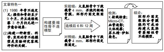
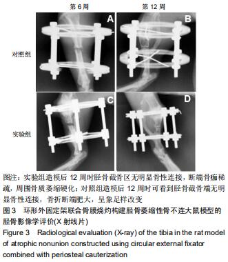
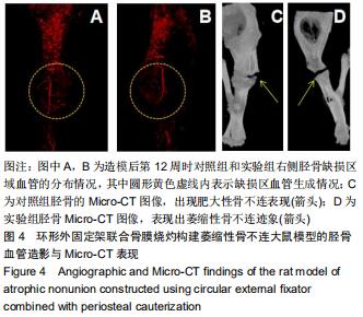
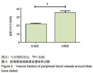
|
[1] EINHORN TA, GERSTENFELD LC. Fracture healing: mechanisms and interventions. Nat Rev Rheumatol. 2015; 11(1):45-54.
[2] KOSTENUIK P, MIRZA FM. Fracture healing physiology and the quest for therapies for delayed healing and nonunion. J Orthop Res. 2017;35(2):213-223.
[3] 胥少汀,葛宝丰,徐印坎.实用骨科学[M].北京:人民军医出版社, 2012:1153-1154.
[4] FONG K, TRUONG V, FOOTE CJ, et al. Predictors of nonunion and reoperation in patients with fractures of the tibia: an observational study. BMC Musculoskelet Disord. 2013;14: 103.
[5] 王海,黄游,何晓青,等.感染性骨不连的治疗现状[J].中国矫形外科杂志,2017,25(5):438-441.
[6] LONG H, ZHU Y, LIN Z, et al. miR-381 modulates human bone mesenchymal stromal cells (BMSCs) osteogenesis via suppressing Wnt signaling pathway during atrophic nonunion development. Cell Death Dis. 2019;10(7):470.
[7] 王亦璁,姜保国.骨与关节损伤[M].北京:人民卫生出版社,2012: 328.
[8] BELL A, TEMPLEMAN D, WEINLEIN JC. Nonunion of the Femur and Tibia: An Update. Orthop Clin North Am. 2016; 47(2):365-375.
[9] RAVEN TF, MOGHADDAM A, ERMISCH C, et al. Use of Masquelet technique in treatment of septic and atrophic fracture nonunion. Injury. 2019;50 Suppl 3:40-54.
[10] 房国军,曲志国,崔正宏,等.大鼠胫骨标准实验性骨不连模型的制作[J].中国组织工程研究,2014,18(18):2795-2800.
[11] 江丽霞,袁瑞娟.骨碎补总黄酮促进骨膜细胞增殖及对兔骨不连的治疗作用[J].中国组织工程研究,2019,23(19):2953-2958.
[12] 梁洪杰.股骨干骨折髓内钉固定术后骨不连原因分析及手术治疗[D].唐山:华北理工大学,2015.
[13] 曾景奇,黄枫,姜自伟,等.一种牵张成骨大鼠实验模型的建立[J].中国实验动物学报,2016,24(1):43-46.
[14] ROBERTO-RODRIGUES M, FERNANDES RM, SENOS R, et al. Novel rat model of nonunion fracture with vascular deficit. Injury. 2015;46(4):649-654.
[15] GÓMEZ-BARRENA E, ROSSET P, LOZANO D, et al. Bone fracture healing: cell therapy in delayed unions and nonunions. Bone. 2015;70:93-101.
[16] GHIASI MS, CHEN J, VAZIRI A, et al. Bone fracture healing in mechanobiological modeling: A review of principles and methods. Bone Rep. 2017;6:87-100.
[17] GRÖNGRÖFT I, WISSING S, MEESTERS DM, et al. Development of a novel murine delayed secondary fracture healing in vivo model using periosteal cauterization. Arch Orthop Trauma Surg. 2019;139(12):1743-1753.
[18] 胥少汀,葛宝丰,徐印坎.实用骨科学[M].北京:人民军医出版社, 2012:464.
[19] 殷建,殷照阳.长骨骨不连的研究进展[J].重庆医科大学学报, 2017,42(2):175-179.
[20] 李德强,李彩霞,李明,等.胫骨萎缩性骨不连骨折局部DSA的初步应用[J].实用放射学杂志,2007(6):781-783.
[21] SUNG MH,徐佳,汪春阳,等.外固定支架在肱骨干萎缩性骨不连中的应用及疗效[J].生物骨科材料与临床研究,2019,16(4): 30-32.
[22] SEN MK, MICLAU T. Autologous iliac crest bone graft: should it still be the gold standard for treating nonunions?. Injury. 2007;38 Suppl 1:S75-S80.
[23] CALORI GM, MAZZA EL, MAZZOLA S, et al. Non-unions. Clin Cases Miner Bone Metab. 2017;14(2):186-188.
[24] 王海生,李长江,张国文,等.附加锁定接骨板结合自体髂骨植骨治疗股骨干骨折交锁髓内钉固定术后骨不连[J].中国骨与关节损伤杂志,2014,29(2):183-184.
[25] 赖丽金,莫浩轩.补骨脂素诱导兔骨内膜间充质干细胞增殖构建细胞支架复合体修复骨不连[J].中国组织工程研究,2020,24(1): 40-44.
[26] ZHANG ZY, TEOH SH, CHONG MS, et al. Neo-vascularization and bone formation mediated by fetal mesenchymal stem cell tissue-engineered bone grafts in critical-size femoral defects. Biomaterials. 2010;31(4): 608-620.
[27] GÓMEZ-BARRENA E, ROSSET P, MÜLLER I, et al. Bone regeneration: stem cell therapies and clinical studies in orthopaedics and traumatology. J Cell Mol Med. 2011; 15(6):1266-1286.
[28] GARCIA P, HISTING T, HOLSTEIN JH, et al. Rodent animal models of delayed bone healing and non-union formation: a comprehensive review. Eur Cell Mater. 2013;26:1-14.
[29] MARCAZZAN S, WEINSTEIN RL, DEL FABBRO M. Efficacy of platelets in bone healing: A systematic review on animal studies. Platelets. 2018;29(4):326-337.
[30] VUOLTEENAHO K, MOILANEN T, MOILANEN E. Non-steroidal anti-inflammatory drugs, cyclooxygenase-2 and the bone healing process. Basic Clin Pharmacol Toxicol. 2008; 102(1):10-14.
[31] MEYERS CA, CASAMITJANA J, CHANG L, et al. Pericytes for Therapeutic Bone Repair. Adv Exp Med Biol. 2018;1109: 21-32.
[32] GARCIA P, HISTING T, HOLSTEIN JH, et al. Rodent animal models of delayed bone healing and non-union formation: a comprehensive review. Eur Cell Mater. 2013;26:1-14.
[33] 潘治军,潘静心,杨振邦,等.二次损伤炎症启动犬萎缩性骨不连模型骨痂生长的组织病理学分析[J].中国骨与关节损伤杂志,2019, 34(10):1041-1045.
[34] 赖丽金,莫浩轩.补骨脂素诱导兔骨内膜间充质干细胞增殖构建细胞支架复合体修复骨不连[J].中国组织工程研究,2020,24(1): 40-44.
[35] 樊涛,郭荣,郑彭.体外冲击波结合骨髓间充质干细胞移植治疗兔骨不连的疗效[J].实用医学杂志,2019,35(18):2874-2877.
[36] 王朋朋. 聚焦式冲击波不同治疗次数对骨不连家兔骨愈合的影响及机制研究[D].蚌埠:蚌埠医学院,2018.
[37] 李月玮.续骨丹外敷疗法对胫骨骨不连大鼠VEGF、TGF-β影响的实验研究[D].南京:南京中医药大学,2018.
[38] 赵子星,李宏宇,席立成,等.体外冲击波疗法联合仙桃草口服用于兔桡骨骨不连临床效果观察[J].山东医药,2016,56(36):31-33.
[39] 林坚平.骨生化标志物(NTX、CTX、OC、BSAP)和QCT骨密度测定在早期诊断兔实验性骨不连的作用[D].广州:南方医科大学,2016.
[40] 李庆虎.骨不连的初步流行病学研究及ADAMTS-7在大鼠骨不连模型中的表达和意义[D].济南:山东大学,2016.
[41] 陈顺有,林然,林清坚.新西兰大白兔桡骨缺损性骨不连模型制作的实验研究[J].福建医药杂志,2015,37(5):54-56.
[42] 喻绍顶,倪卫东.体外冲击波联合增强型纤维蛋白胶负载骨生长因子治疗兔骨不连的研究[J].中国免疫学杂志,2015,31(9): 1186-1190+1194.
[43] 刘宇强,李平,赵爱玲.不同时间冲击波干预对骨不连家兔成骨细胞VEGF的影响[J].中华临床医师杂志(电子版),2015,9(9): 1658-1663.
[44] 申震,姜自伟,李定,等.基于Masquelet诱导膜技术比较不同固定方式构建的胫骨大段骨缺损模型[J].中国实验动物学报,2018, 26(6):673-680.
[45] 娄盛涵,张里程,唐佩福.Ilizarov技术治疗骨不连的研究进展[J].解放军医学院学报,2016,37(12):1308-1311.
[46] ZHANG Z, SWANSON WB, WANG YH, et al. Infection-free rates and Sequelae predict factors in bone transportation for infected tibia: a systematic review and meta-analysis. BMC Musculoskelet Disord. 2018;19(1):442.
|