中国组织工程研究 ›› 2021, Vol. 25 ›› Issue (21): 3349-3353.doi: 10.3969/j.issn.2095-4344.3850

• 骨与关节图像与影像 bone and joint imaging • 上一篇    下一篇

术中画线法与三维重建术前模拟测量股骨截骨旋转角度对比的体外实验

林天烨1,杨  鹏1,熊冰朗2,何晓铭2,颜新昊2,张  进3,何  伟1,魏秋实1   

  1. 1广州中医药大学第三附属医院,广东省广州市   510405;2广州中医药大学第一临床医学院,广东省广州市   510080;3广州中医药大学,广东省广州市   510000
  • 收稿日期:2020-07-14 修回日期:2020-07-16 接受日期:2020-08-26 出版日期:2021-07-28 发布日期:2021-01-23
  • 通讯作者: 魏秋实,副主任医师,博士后,博士生导师,广州中医药大学第三附属医院,广东省广州市 510405
  • 作者简介:林天烨,男,1992年生,广东省云浮市人,在读博士,主要从事中西医结合治疗骨伤科疾病研究。
  • 基金资助:
    国家自然科学基金(81873327),项目负责人:何伟;广东省中医药局科研项目(20191116),项目负责人:杨鹏

Comparison of preoperative three-dimensional reconstruction simulation and intraoperative drawing of femoral osteotomy to measure rotation angle in vitro 

Lin Tianye1, Yang Peng1, Xiong Binglang2, He Xiaoming2, Yan Xinhao2, Zhang Jin3, He Wei1, Wei Qiushi1    

  1. 1Third Affiliated Hospital of Guangzhou University of Chinese Medicine, Guangzhou 510405, Guangdong Province, China; 2First Affiliated Hospital of Guangzhou University of Chinese Medicine, Guangzhou 510080, Guangdong Province, China; 3Guangzhou University of Chinese Medicine, Guangzhou 510000, Guangdong Province, China
  • Received:2020-07-14 Revised:2020-07-16 Accepted:2020-08-26 Online:2021-07-28 Published:2021-01-23
  • Contact: Wei Qiushi, Associate chief physician, MD, Doctoral supervisor, Third Affiliated Hospital of Guangzhou University of Chinese Medicine, Guangzhou 510405, Guangdong Province, China
  • About author:Lin Tianye, Doctoral candidate, Third Affiliated Hospital of Guangzhou University of Chinese Medicine, Guangzhou 510405, Guangdong Province, China
  • Supported by:
    the National Natural Science Foundation of China, No. 81873327 (to HW); the Scientific Research Project of Guangdong Provincial Bureau of Traditional Chinese Medicine, No. 20191116 (to YP)

摘要:

文题释义:
股骨旋转截骨术:用于髋关节脱位的手术治疗,通过股骨旋转截骨可矫治严重股骨颈前倾畸形,同时通过股骨缩短截骨可使股骨头在无张力下复位,还可做楔形截骨矫正髋外翻或髋内翻畸形。
术前可视化模拟:术前利用计算机图形学和图像处理技术将数据转换成图像在屏幕上显示出来,CT扫描得到的DICOM数据分别导入到Mimics 16.0系统、Geomagic-Studio11系统进行三维重建,通过Solidworks进行术前模拟。

背景:临床上治疗发育性髋关节发育不良股骨颈前倾角增大者常通过股骨上端旋转截骨来实现,而如何术中精准控制旋转角度及位移是手术成功的关键。
目的:对比三维重建术前模拟与术中画线法两种股骨截骨旋转角度测量方法在矫正股骨颈前倾角中的价值。
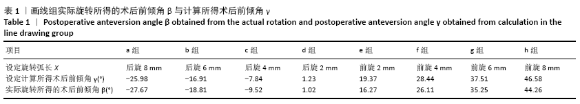
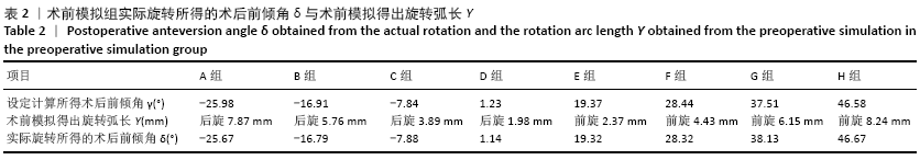
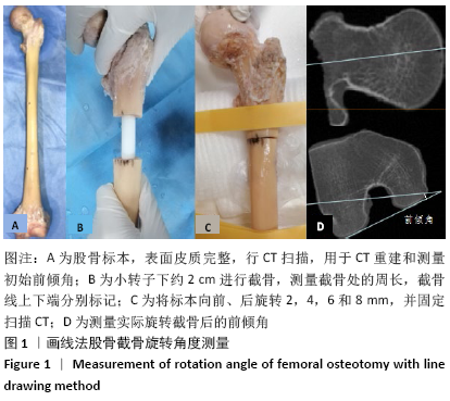
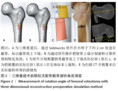
方法:取正常成人股骨标本进行体外旋转截骨,画线组通过公式计算股骨上端向前及向后各旋转2,4,6,8 mm所得的前倾角(共8组),然后在股骨标本上完成旋转截骨操作,CT扫描测量术后实际所得前倾角;术前模拟组对完整股骨标本行三维重建,对上述计算所得前倾角,通过Solidworks进行术前模拟股骨截骨旋转至特定角度,测量股骨截骨端表面位移(弧长),然后根据弧长在股骨标本上进行旋转截骨,CT扫描测量术后实际所得前倾角。将两组术后实际所得股骨颈前倾角与计算所得股骨颈前倾角进行对比分析。
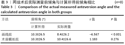
结果与结论:①画线组术后实际测量的前倾角为(8.4±26.2)°,术前计算所得前倾角为(10.3±26.5)°,两者比较差异有显著性意义(P < 0.05);②术前模拟组术后实际测量的前倾角为(10.4±26.6)°,与术前计算所得前倾角(10.3±26.5)°比较差异无显著性意义(P > 0.05);③结果表明,三维重建术前模拟与术中画线法指导股骨旋转截骨均能有效纠正过大股骨前倾角,三维重建术前模拟较术中画线法的误差更小,三维重建术前模拟可以使股骨旋转截骨更加精准化、个性化、智能化。

关键词: 骨, 画线法, 发育性髋关节发育不良, 股骨截骨, 前倾角, 脱位, 三维CT, 术前模拟

Abstract: BACKGROUND: Clinical treatment of the increased anteversion angle of the femoral neck in developmental dysplasia of the hip is often achieved by rotating osteotomy at the upper end of the femur, and how to accurately control the rotation angle and displacement during the operation is the key to the success of the operation.
OBJECTIVE: To compare the value of two femoral osteotomy rotation angle measurement methods in the correction of the anteversion angle of the femoral neck by three-dimensional reconstruction simulation before operation and intraoperative line drawing.
METHODS: Normal adult femur specimens were taken for external rotation osteotomy. In the line drawing group, formulas were used to calculate the anterior inclination angle of the upper end of the femur by 2, 4, 6, and 8 mm (total 8 groups). The rotation osteotomy operation was completed. CT scan was used to measure the actual postoperative anteversion angle. In the preoperative simulation group, three-dimensional reconstruction of the intact femoral specimen was performed, and the calculated anteversion angle was performed by Solidworks to simulate the femoral osteotomy rotation to a specific angle before surgery. The surface displacement (arc length) of the femoral osteotomy end was measured, and then a rotary osteotomy was performed on the femoral specimen according to the arc length. The actual anteversion angle obtained after the CT scan was measured. The actual postoperative femoral neck anteversion angle and the calculated femoral neck anteversion angle were compared and analyzed in both groups.
RESULTS AND CONCLUSION: (1) The actual measured anteversion angle after the line drawing group was (8.4±26.2)°, and the calculated preoperative anteversion angle was (10.3±26.5)°; there was a significant difference between the two (P < 0.05). (2) The actual measured anteversion angle in the preoperative simulation group was (10.4±26.6)°, which was not significantly different from the calculated preoperative anteversion angle (10.3±26.5)° (P > 0.05). (3) The results showed that three-dimensional reconstruction pre-simulation and intraoperative line-drawing method to guide femoral rotation osteotomy can effectively correct the excessive femoral anteversion angle. The preoperative simulation of three-dimensional reconstruction has smaller errors than the intraoperative line-drawing method. The preoperative simulation of three-dimensional reconstruction can make the femoral rotation osteotomy more precise, personalized and intelligent.   

Key words: bone, line drawing method, developmental hip dysplasia, femoral osteotomy, anteversion, dislocation, three-dimensional CT, preoperative simulation

中图分类号: