中国组织工程研究 ›› 2020, Vol. 24 ›› Issue (6): 843-849.doi: 10.3969/j.issn.2095-4344.2424

• 骨科植入物 orthopedic implant • 上一篇    下一篇

微型锁定接骨板与外固定支架治疗骰骨粉碎骨折的比较

王  敏,马文泽,李文成,梁  材,王进辉   

  1. 天津港口医院骨科,天津市  300456
  • 收稿日期:2019-04-22 修回日期:2019-04-29 接受日期:2019-07-05 出版日期:2020-02-28 发布日期:2020-01-17
  • 作者简介:王敏,男,1984年生,山西省保德县人,汉族,2009年山西医科大学毕业,硕士,主治医师,主要从事足踝及运动损伤方面的研究。
  • 基金资助:
    天津市滨海新区卫生计生委科研基金青年项目(2018BWKQ030)

A comparative study of mini-locking plate and external fixator in the treatment of comminuted cuboid fracture

Wang Min, Ma Wenze, Li Wencheng, Liang Cai, Wang Jinhui   

  1. Department of Orthopedics, Tianjin Port Hospital, Tianjin 300456, China
  • Received:2019-04-22 Revised:2019-04-29 Accepted:2019-07-05 Online:2020-02-28 Published:2020-01-17
  • About author:Wang Min, Master, Attending physician, Department of Orthopedics, Tianjin Port Hospital, Tianjin 300456, China
  • Supported by:
    the Scientific Research Fund Youth Project of Tianjin Binhai New Area Health and Family Planning Commission, No. 2018BWKQ030

摘要:

文题释义:
中足外侧柱:根据Myerson中足三柱理论,中足外侧柱包括骰骨、跟骰关节、跖骰关节,其中骰骨是中足唯一支撑外侧柱的骨头,几乎参与足的所有内在运动。
足外侧柱活动度:跟骰关节几乎没有运动,外侧柱的运动全部发生在骰骨关节面远端,跖跗外侧关节的活动范围是跖跗内侧关节的3倍。

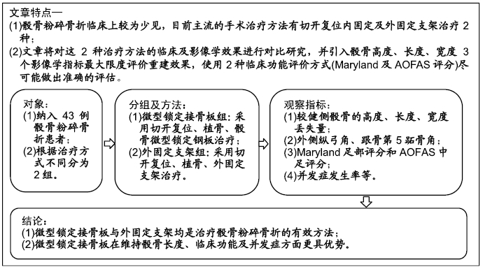
背景:骰骨粉碎骨折的手术治疗方式主要有切开复位内固定及外固定治疗2种,随着内固定器械的发展,尤其是微型锁定接骨板的出现改善了内固定的治疗效果。

目的:比较微型锁定接骨板与外固定支架治疗骰骨粉碎骨折的临床及影像学效果。

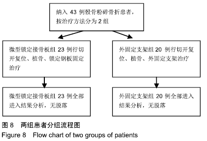
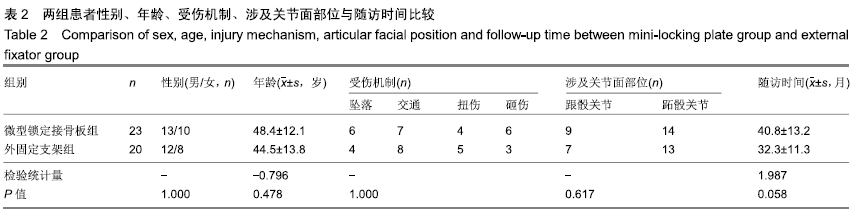
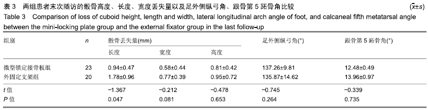
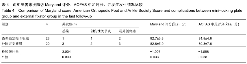
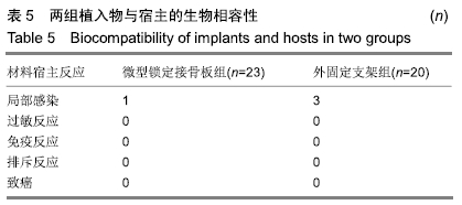
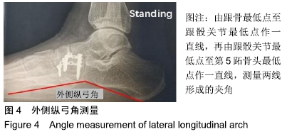
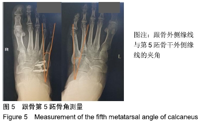
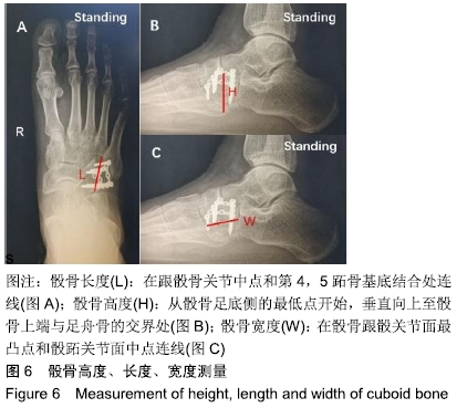
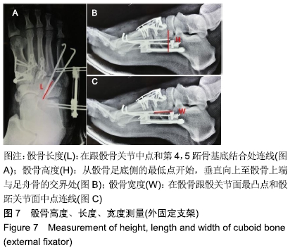
方法:回顾性分析2013年1月至2018年3月天津港口医院骨科收治的43例骰骨粉碎骨折患者资料,根据治疗方式不同分为2组,微型锁定接骨板组23例,外固定支架组20例。2组患者对治疗方案均知情同意,且得到医院伦理委员会批准。2组患者术前均行足部CT及三维重建扫描证实为骰骨粉碎骨折,术中均进行了异体骨的植骨支撑,外固定支架于术后三四个月内去除。末次随访时行双足站立正侧位X射线检查,比较2组患者较健侧骰骨的高度、长度、宽度丢失量,以及外侧纵弓角、跟骨第五跖骨角、Maryland足部评分、美国足踝外科协会中足评分、并发症发生率等。

结果与结论:①所有患者均获得1年以上随访;②2组患者末次随访骰骨高度丢失量、宽度丢失量、末次外侧纵弓角、跟骨第5跖骨角比较差异无显著性意义(P > 0.05);③2组患者在骰骨长度丢失量、Maryland足部评分、美国足踝外科协会中足评分、并发症发生率方面比较,差异均有显著性意义(P < 0.05),微型锁定接骨板组优于外固定支架组;④提示微型锁定接骨板与外固定支架均是治疗骰骨粉碎骨折的有效方法。微型锁定接骨板在维持骰骨长度、临床功能及减少并发症方面更具优势,随着时间的推移可获得足部稳定、功能良好的结果。

ORCID: 0000-0003-2285-2223(王敏)

中国组织工程研究杂志出版内容重点:人工关节;骨植入物;脊柱骨折;内固定;数字化骨科;组织工程

关键词: 骰骨骨折, 微型锁定接骨板, 外固定支架, 足外侧柱, 创伤性关节炎, 并发症

Abstract:

BACKGROUND: Open reduction internal fixation and external fixation are two main surgical treatments for comminuted cuboid fractures. With the development of internal fixation devices, especially the appearance of mini-locking plate, the therapeutic effect of internal fixation has been improved.        

OBJECTIVE: To compare the clinical and imaging effects of mini-locking plate and external fixator in the treatment of comminuted cuboid fracture.          

METHODS: The data of 43 patients with comminuted cuboid fracture admitted to the Department of Orthopedics, Tianjin Port Hospital from January 2013 to March 2018 were retrospectively analyzed. According to different treatment methods, the patients were divided into two groups: 23 cases in the mini-locking plate group and 20 cases in the external fixator group. All patients signed the informed consent. This study was approved by the Hospital Ethics Committee. The patients in both groups were confirmed as comminuted cuboid fracture by CT and three-dimensional reconstruction scan before operation. Bone graft support of allograft was performed during the operation, and the external fixator was removed within 3-4 months after operation. At the last follow-up, bilateral standing and anteroposterior and lateral X-ray examination was performed to compare the loss of height, length, width, lateral longitudinal arch angle, calcaneal fifth metatarsal angle, Maryland foot score, American Orthopedic Foot and Ankle Society Score and the incidence of complications between the two groups. 

RESULTS AND CONCLUSION: (1) All patients were followed up for more than one year. (2) There were no significant differences in height loss of cuboid bone, width loss, last lateral longitudinal arch angle, and calcaneal fifth metatarsal angle between the two groups (P > 0.05). (3) There were significant differences in the loss of cuboid bone length, Maryland foot score and American Orthopedic Foot and Ankle Society Score and the incidence of complications between the two groups (< 0.05). Mini-locking plate group was superior to the external fixator group in above indexes. (4) These findings indicated that mini-locking plate and external fixator are effective methods for the treatment of comminuted cuboid fracture. Micro-locking plate has more advantages in maintaining the length of cuboid bone, clinical function and reducing complications. With the prolongation of time, stable foot and good function can be achieved.           

Key words: cuboid fracture, mini-locking plate, external fixator, lateral column of foot, traumatic arthritis, complications

中图分类号: