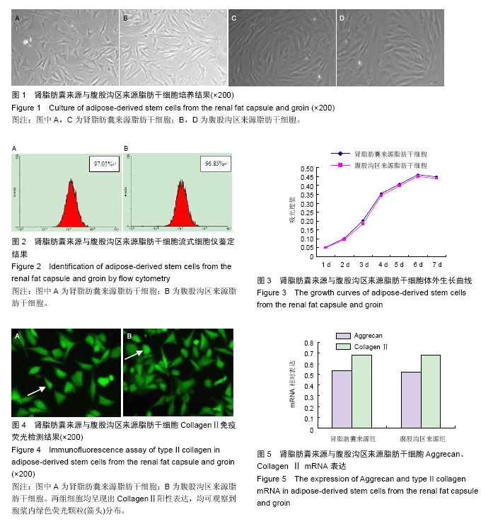
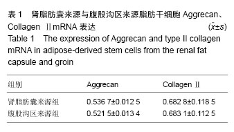
| [1] 张慧,郑红光,张德伟,等.雌激素影响冻存肾脂肪囊来源脂肪间充质干细胞的成脂分化[J].中国组织工程研究,2013, 17(27):4998-5004.
[2] 常春娟,贾赤宇,胡永亮,等.大鼠肾脂肪囊来源干细胞修复深Ⅱ°烫伤创面的实验研究[C].中国康复医学会修复重建外科专业委员会第19次学术交流会暨中国医师协会烧伤科医师分会2012年年会论文集,2012:357.
[3] 常春娟,贾赤宇,田甜,等.大鼠肾脂肪囊来源干细胞修复深Ⅱ°烫伤创面的实验研究[C].第八届全国创伤修复(愈合)与组织再生学术交流会论文集,2012:190.
[4] 訾利影,郑红光,张德伟,等.氯甲基苯甲酰胺标记大鼠肾脂肪囊来源脂肪间充质干细胞的可行性[J].中国组织工程研究,2012,16(19):3520-3524.
[5] 付勤,王勇,于冬冬,等.外源性TGF-β1及IGF-1诱导大鼠脂肪干细胞向软骨细胞分化的实验研究[J].中国骨质疏松杂志,2007,13(12):848-853.
[6] 王勇.外源性TGF-β1及IGF-1诱导大鼠脂肪干细胞向软骨细胞分化的实验研究[D].沈阳:中国医科大学,2008.
[7] 柴仪.BMP-9通过BMP信号通路诱导鼠脂肪源性干细胞分化为成骨细胞及经皮椎体后凸成形术治疗高龄骨质疏松性椎体压缩骨折及并发症的防治[D].石家庄:河北医科大学,2013.
[8] 吴畏,郑红光,张德伟,等.肾脂肪囊来源脂肪千细胞的体外培养及其生物学特性的研究[J].细胞与分子免疫学杂志, 2010,26(11):1078-1081.
[9] Kølle SF, Fischer-Nielsen A, Mathiasen AB, et al. Enrichment of autologous fat grafts with ex-vivo expanded adipose tissue-derived stem cells for graft survival: a randomised placebo-controlled trial. Lancet. 2013;382(9898):1113-1120.
[10] 赵建辉,李龙,梁丽华,等.人与家兔脂肪来源干细胞体外培养特性的比较[J].中国组织工程研究,2012,16(14):2540- 2544.
[11] Koh KS, Oh TS, Kim H, et al. Clinical application of human adipose tissue-derived mesenchymal stem cells in progressive hemifacial atrophy (Parry-Romberg disease) with microfat grafting techniques using 3-dimensional computed tomography and 3-dimensional camera. nn Plast Surg. 2012;69(3): 331-337.
[12] 江波,廖毅,童庭辉,等.脂肪来源干细胞诱导表皮细胞化的研究现状与进展[J].中国组织工程研究与临床康复,2011, 15(27):5104-5107.
[13] Han DS, Chang HK, Kim KR, et al. Consideration of bone regeneration effect of stem cells: comparison of bone regeneration between bone marrow stem cells and adipose-derived stem cells. J Craniofac Surg. 2014;25(1):196-201.
[14] 李力群,高建华,鲁峰,等.人脂肪来源干细胞与Ⅰ型胶原支架体外复合培养的研究[J].中华实验外科杂志,2012, 29(3):421-423.
[15] Derby BM, Dai H, Reichensperger J, et al. Adipose-derived stem cell to epithelial stem cell transdifferentiation: a mechanism to potentially improve understanding of fat grafting's impact on skin rejuvenation.Aesthet Surg J. 2014;34(1):142-153.
[16] 杨杰,郭能强,孙家明,等.体外诱导乳房内脂肪来源干细胞向上皮细胞分化的实验研究[J].中华整形外科杂志,2014, 30(3):209-214.
[17] Aarum J, Sandberg K, Haeberlein SL, et al. Migration and differentiation of neural precursor cells can be directed by microglia. Proc Natl Acad Sci U S A. 2003; 100(26):15983-15988.
[18] Narazaki G, Uosaki H, Teranishi M, et al. Directed and systematic differentiation of cardiovascular cells from mouse induced pluripotent stem cells. Circulation. 2008;118(5):498-506.
[19] Otsu M, Sai T, Nakayama T, et al. Uni-directional differentiation of mouse embryonic stem cells into neurons by the neural stem sphere method. Neurosci Res. 2011;69(4):314-321.
[20] 徐扬阳,姜南,杨柳,等.重组人碱性成纤维细胞生长因子对人脂肪干细胞分化为脂肪细胞的影响[J].中华医学美学美容杂志,2013,19(2):134-137.
[21] Vergroesen PP, Kroeze RJ, Helder MN, et al. The use of poly(L-lactide-co-caprolactone) as a scaffold for adipose stem cells in bone tissue engineering: application in a spinal fusion model. Macromol Biosci. 2011;11(6):722-730.
[22] 陈冲,闫俊灵,李梁,等.人脂肪来源间充质干细胞的制备及其质量检验方法[J].武警医学,2015,26(2):170-174.
[23] 刘学晖,郭澍.脂肪来源干细胞提取、分离、免疫表型及分化的研究进展[J].中国美容整形外科杂志,2013,24(2): 112-115.
[24] Gentile P, De Angelis B, Pasin M, et al. Adipose-derived stromal vascular fraction cells and platelet-rich plasma: basic and clinical evaluation for cell-based therapies in patients with scars on the face. J Craniofac Surg. 2014;25(1):267-272.
[25] Kim YJ, Hwang SH, Cho HH, et al. MicroRNA 21 regulates the proliferation of human adipose tissue- derived mesenchymal stem cells and high-fat diet- induced obesity alters microRNA 21 expression in white adipose tissues. J Cell Physiol. 2012;227(1): 183-193.
[26] 郑木平,曾云霞,魏波,等.脂肪干细胞向软骨分化构软骨复合体和移植界面的实验研究[J].牡丹江医学院学报,2014, 35(4):8-12.
[27] Marappagounder D, Somasundaram I, Dorairaj S, et al. Differentiation of mesenchymal stem cells derived from human bone marrow and subcutaneous adipose tissue into pancreatic islet-like clusters in vitro. Cell Mol Biol Lett. 2013;18(1):75-88.
[28] 安荣泽,赵俊延,王兆杰,等.脂肪干细胞与骨髓间充质干细胞成软骨能力的比较[J].中国组织工程研究,2013,17(32): 5793-5798.
[29] 王和庚,黎洪棉,崔世恩,等.带血管肌筋膜包埋脂肪干细胞载体复合物构建血管化组织工程脂肪[J].中国组织工程研究,2013,17(18):124-132.
[30] Lee RH, Kim B, Choi I, et al. Characterization and expression analysis of mesenchymal stem cells from human bone marrow and adipose tissue. Cell Physiol Biochem. 2004;14(4-6):311-324.
[31] Kim WS, Park BS, Kim HK, et al. Evidence supporting antioxidant action of adipose-derived stem cells: protection of human dermal fibroblasts from oxidative stress.J Dermatol Sci. 2008;49(2):133-142.
[32] 陈亮,吴柏霖,吴明珑,等.解剖部位差异对脂肪干细胞生物学行为及成骨分化能力的影响[J].华中科技大学学报:医学版,2013,42(5):564-568.
[33] Toyoda M, Matsubara Y, Lin K, et al. Characterization and comparison of adipose tissue-derived cells from human subcutaneous and omental adipose tissues. Cell Biochem Funct. 2009;27(7):440-447.
[34] Kang SW, Do HJ, Han IB, et al. Increase of chondrogenic potentials in adipose-derived stromal cells by co-delivery of type I and type II TGFβ receptors encoding bicistronic vector system. J Control Release. 2012;160(3):577-582.
[35] 刘元媛,柳大烈,南华,等.Choukroun's PRF对体外培养人脂肪干细胞增殖及成骨分化的影响[J].中国美容医学, 2013,22(1):40-43.
[36] 察鹏飞,高建华,陈阳,等.人细胞外基质支架联合脂肪干细胞构建脂肪组织[J].中华实验外科杂志,2012,29(6): 1085-1088.
[37] Alharbi Z, Opländer C, Almakadi S, et al. Conventional vs. micro-fat harvesting: how fat harvesting technique affects tissue-engineering approaches using adipose tissue-derived stem/stromal cells. J Plast Reconstr Aesthet Surg. 2013;66(9):1271-1278.
[38] 范开防,王国栋,刘兴龙,等.人上睑眶隔脂肪来源的脂肪干细胞的分离培养及鉴定[J].现代医药卫生,2012,28(19): 2881-2882,2885.
[39] 察鹏飞,高建华,陈阳,等.体外碱性成纤维细胞生长因子聚乳酸纳米缓释微球对人脂肪干细胞增殖和成脂分化的影响[J].中华医学美学美容杂志,2012,18(2):132-135.
[40] 赵宝成,赵博,顾蓓,等.猪脂肪来源干细胞分离、培养及多向诱导分化研究[J].中华实验外科杂志,2011,28(7): 1099-1101. |
.jpg)


.jpg)