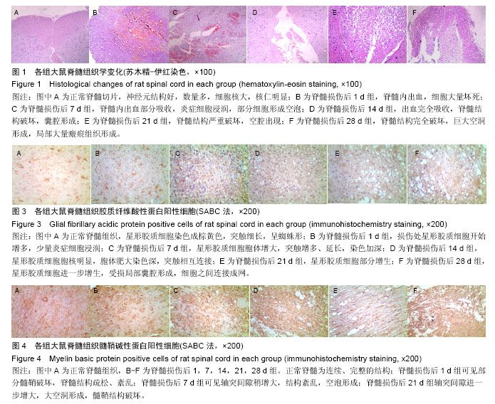
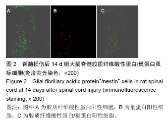
| [1] Sofroniew MV, Vinters HV. Astrocytes:biology and pathology. Acta Neuropathologica, 2010;119(1):7-35.
[2] Matsuura I, Taniguchi J, Hata K, et al. BMP inhibition enhances axonal growth and functional recovery after apinal cord injury. J Neurochem.2008;105(4):1471-1479.
[3] 胡凌云,张建英,林宏,等.脊髓损伤模型大鼠星形胶质化形成与Akt/mTOR/p70S6K信号通路的激活[J].中国组织工程研究,2015,19(5):697-703.
[4] 李珂珂,宗少晖.脊髓损伤中的免疫炎性细胞因子[J].中国组织工程研究,2015,19(53):8646.
[5] Rolls A,Shcchter R,Schwartz M. The bright side of the glialscar in CNS repair. Nat Rev Neurosci. 2009;10(3): 235-241.
[6] Natarajan R,Singal V,Benes R,et al.STAT3 modulation to enhance motor neuron differentiantion in human neural stem cells.PLOS One.2014;9(6):e100405.
[7] Hong P,Jiang M,Li H.Functional requirement of dicerland miR-17-5p in reactive astrocyte proliferation after spinal cord injury in the mouse. Glia. 2014;62(12): 2044-2060.
[8] Qian DX,Zhang HT,Cai YQ,et al.Expression of tyrosine kinase receptor C in the segments of the spinal cord and the cerebral cortex after cord transection in adult rats.Neurosci Bull.2011;27(2):83-90.
[9] 刘静,韩冬梅,王志东,等.脐带间充质干细胞治疗脊髓损伤临床分析[J].中华损伤与修复杂志(电子版),2011,6(4): 564-569.
[10] 杨平林,贺西京,李浩鹏,等.成年大鼠脊髓损伤后反应性星形胶质细胞增生和巢蛋白表达的相关性及意义[J].南方医科大学学报,2008,28(10):1752-1755.
[11] Buffo A,Rite I,Tripathi P,et al.Origin and progeny of reactive gliosis: A source of multipotent cells in the injured brain.Proc Natl Acad Sci USA.2008;105(9):3581-3586.
[12] Yang H,Cheng XP,Li JW,et al.De-differentiation response of cultured astrocytes to injury induced by scratch or conditioned culture medium of scratch-insulted astrocytes.Cell Mol Neutobiol.2010;29(4):445-473.
[13] Gris P,Tighe A,Levin D,et al.Transcriptional regulation of scar gene expression in primary astrocytes.Glia.2007;55(11):1145-55.
[14] Busch SA,Horn KP,Cuascut FX,et al.Adult NG cells are permissive to neurite outgrowth and stabilize sensory axons during macrophage-induced axonal dieback after spinal cord injury.J Neurosic.2010;30(1):255-265.
[15] Nawashiro H,Messing A,Azzam N.Mice lacking GFAP are hrpersensitive to traumatic cerebro-spinal injury. Neuroreport.1998;9(8):1691-1696.
[16] Okano H,Kaneko S,Okada S,et al.Regeneration-based therapies for spinal cord injuries.Neurochem Int.2007; 51(2-4):68-73.
[17] Pekny M,Johansson CB,Eliasson C,et al.Abnormal reaction to central nervous system injury in mice lacking glial fibrillary acidic protein and vimentin.J cell Biol.1999;(3):503-514.
[18] Liesi P,Kauppila T.Induction of type IV collagen and other basement membrane associated proteins after apinal cord injury of the adult rat participate in formation of the glia scar.Exp Neurol.2002;173(7):31-45.
[19] Shimizu F,Sano Y,Saito K,et al.Pericyte-derived glial cell line-derived neurotrophic factor increase the expression of claudin-5 in the blood-brain barrier and the blood-nerve barrier.Neurochem Res.2012;37(2):401-409.
[20] Sofroniew MV.Reactive astrocytes in neural repair and protection .Neuroscientist.2005;11(5):400-407.
[21] Potokar M,Stenovec M,Jorgacevski J,et al.Regulation of AQP4 surface expression via vesicle mobility in astrocytes.Glia, 2013;61(6):917-928.
[22] 邹云,余资江,林金棠.大鼠脊髓半横断损伤后胶质细胞反应性增生的变化规律及意义[J].贵阳医学院学报, 2015, 40(9):905-909.
[23] Yamazaki Y,Yada K,Morii S, et al. Diagnostic significance of serum neuro-specific endase and myelin basic protein assay in patients with acute head injury.Surg Neurol.1995;(3):267-269.
[24] Su Z,Yuan Y,Chen J,et al.Reactive astrocytes inhibit the survival and differentiation of oligodendrocyte precursor cells by secreted TNF-alpha.J Neurotrauma. 2011;28(6):1089-1100.
[25] Toyooka T,Nawashiro H,Shinomiya N,et al. Down-regulation of glial fibrillary acidic protein and vimentin by RNA interference improves acute urinary dysfunction associated with spinal cord injury in rats.J Neurotrauma.2011;28(4):607-618.
[26] Leung YK,Pankhurst M,Dunlop SA,et al. Metallothionein induces a regenerative reative astrocyte phenotype via JAK/STAT and RhoA signaling pathways.Exp Neurol.2010;221(1):98-106.
[27] Fisher D,Xing B,Dill J,et al. Leukocyte common antigenrelated phosphatase is a functional receptor fo chondroitin sulfate proteoglycan axon growth inhibitors.J Neurosci.2011;31(40):14051-14066.
[28] Fry EJ,Chagnon MJ,Lopez-Vales R,et al.Corticospinal tract regeneration after spinal cord injury in recptor protein tyrosine phosphatase sigma deficine mice. Glia. 2010;58(4):423-433.
[29] 12Kang W,Hebert JM.Signaling pathways in reactive astrocytes: a genetic perspective.Mol Neurobiol. 2011; 43(3):147-154. |
.jpg)


.jpg)