中国组织工程研究 ›› 2016, Vol. 20 ›› Issue (40): 5973-5978.doi: 10.3969/j.issn.2095-4344.2016.40.007

• 脑及脊髓损伤动物模型 Animal models of brain and spinal cord injuries • 上一篇    下一篇

急性马尾神经压迫模型的脊髓组织病理变化

王 展1,李浩鹏2,贺西京2,郝定均3,张 堃1,陈明霞4,雷 霆5   

  1. 西安交通大学医学院附属红会医院,1创伤骨科,3脊柱科,陕西省西安市 710054;2西安交通大学第二临床学院骨二科,陕西省西安市 710003;西安交通大学医学部,4电镜室,5病理教研室,陕西省西安市 710061
  • 修回日期:2016-08-07 出版日期:2016-09-30 发布日期:2016-09-30
  • 作者简介:王展,男,1970年生,陕西省西安市人,汉族,硕士,副主任医师,主要从事脊柱与脊髓损伤,外伤后骨髓炎治疗,外伤后畸形矫正研究。
  • 基金资助:

    国家自然科学基金面上资助项目(30271310)

Pathological changes in the spinal cord of a model of acute cauda equina compression

Wang Zhan1, Li Hao-peng2, He Xi-jing2, Hao Ding-jun3, Zhang Kun1, Chen Ming-xia4, Lei Ting5   

  1. 1Department of Orthopedic Trauma, 3Department of Spine, Xi’an Honghui Hospital, Health Science Center, Xi’an Jiaotong University, Xi’an 710054, Shaanxi Province, China; 2Second Department of Orthopedics, the Second Clinical College of Xi’an Jiaotong University, Xi’an 710003, Shaanxi Province, China; 4Room of Electron Microscope, 5Department of Pathology, Health Science Center, Xi’an Jiaotong University, Xi’an 710061, Shaanxi Province, China
  • Revised:2016-08-07 Online:2016-09-30 Published:2016-09-30
  • About author:Wang Zhan, Master, Associate chief physician, Department of Orthopedic Trauma, Xi’an Honghui Hospital, Health Science Center, Xi’an Jiaotong University, Xi’an 710054, Shaanxi Province, China
  • Supported by:

    the National Natural Science Foundation of China, No. 30271310

摘要:

文章快速阅读:


文题释义:

马尾神经:在脊髓圆锥以下的腰骶神经根称为马尾,由L2-5、S1-5及尾节发出的共10对神经根组成。
马尾神经综合征:由腰骶部骨性结构改变或/和软组织压迫等因素引起,表现为膝关节平面肢体运动、感觉、痛觉、知觉、温度功能障碍,如神经缺血时间过长或并发继发水肿麻痹加重,会导致截瘫。
 
摘要
背景:动物实验发现,压迫马尾神经可导致腰骶段脊髓前角运动神经元发生凋亡。
目的:观察急性马尾神经压迫后家犬相应脊髓节段组织的病理变化。
方法:将27只犬随机分为9组,每组3组,其中1组为正常对照,7个实验组在L6椎板下硬膜外脂肪上方置入空水囊,进行注水压迫,压迫时间分别设置为4,8,12,24,48,72,168 h;假手术组只置入空水囊,不进行注水压迫。达到压迫时间后,取马尾神经发出段脊髓及相邻头端脊髓,进行组织病理学检查。
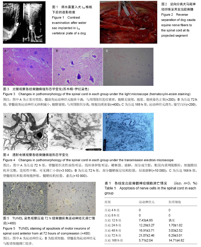
结果与结论:①光镜观察结果:压迫4-48 h,脊髓前角前运动神经元未见明显改变;压迫72 h后,观察到运动神经元体积减小,细胞膜皱缩,与周围组织分离,细胞均质浓染;压迫168 h可见运动神经元消失,但相邻头端脊髓切片未见脊髓前角运动神经元形态异常;②电镜观察结果:压迫12 h,脊髓组织开始肿胀,随压迫时间的延长,脊髓组织肿胀加重,胶质细胞肿胀明显,至168 h时髓鞘板层结构溶解,轴突空泡化,轴浆外溢,有炎性损伤特征样改变;③前角运动神经元凋亡检测结果:压迫12 h开始出现凋亡,随后逐渐增多,至168 h仍呈增高趋势。
中国组织工程研究杂志出版内容重点:肾移植肝移植移植;心脏移植;组织移植;皮肤移植;皮瓣移植;血管移植;器官移植组织工程

ORCID:
0000-0002-8010-455X(王展)

关键词: 实验动物, 神经损伤与修复动物模型, 马尾神经, 急性压迫, 脊髓组织, 凋亡, 继发性炎症, 国家自然科学基金

Abstract:

BACKGROUND: Animal studies have shown that cauda equina compression can induce apoptosis of lumbosacral spinal cord anterior horn motor neurons.

OBJECTIVE: To explore the pathological change in lumbosacral spinal cord after acute cauda equina compression in dogs.
METHODS: A total of 27 dogs were randomly divided into nine groups, with three dogs in each group. There were one normal control group, seven experimental groups and one sham surgery group. In the experimental group, an empty water sac was implanted above epidural fat below L6 vertebral plate. Compression was given by injecting water at 4, 8, 12, 24, 48, 72 and 168 hours. In the sham surgery group, an empty water sac was implanted, but compression was not given. At the time of compression, the spinal cord sent out by cauda equina nerve and adjacent to the head end was subjected to histopathological examination. 
RESULTS AND CONCLUSION: (1) Results of light microscope: at 4-48 hours of compression, spinal cord anterior horn motor neurons did not alter. At 72 hours, motor neurons became small, cell membrane shrank and separated from surrounding tissues. Cells were homogenous and darkly stained. At 168 hours, motor neurons disappeared, but spinal cord sections of the adjacent head end did not shown abnormal motor neurons in the spinal cord anterior horn. (2) Results of electron microscope: at 12 hours, spinal cord tissue began to swell, and the swelling aggravated with prolonged time of compression. The swelling of glial cells was apparent. At 168 hours, myelin sheath structure dissolved; axons showed vacuolization; axoplasm spilled, and exhibited inflammatory injury-like changes. (3) Apoptotic results of spinal cord anterior horn motor neurons: apoptosis appeared at 12 hours of compression, became increased, and showed an increased trend at 168 hours.

中国组织工程研究杂志出版内容重点:肾移植肝移植移植;心脏移植;组织移植;皮肤移植;皮瓣移植;血管移植;器官移植组织工程

Key words:  Models, Animal, Cauda Equina, Spinal Cord, Tissue Engineering

中图分类号: