中国组织工程研究 ›› 2016, Vol. 20 ›› Issue (24): 3609-3614.doi: 10.3969/j.issn.2095-4344.2016.24.017
• 肌肉肌腱韧带组织构建 tissue construction of the muscle, tendon and ligament • 上一篇 下一篇
高频超声检查成人足背侧Lisfranc韧带的图像特征
陈佩瑾,玛依努尔•买买提明,侯 霞,张爱红,刘纯红
- 新疆医科大学第二附属医院超声科,新疆维吾尔自治区乌鲁木齐市 830063
High frequency ultrasonic imaging of the dorsal Lisfranc ligament in the adults
Chen Pei-jin, Mayinuer Maimaitiming, Hou Xia, Zhang Ai-hong, Liu Chun-hong
- Department of Ultrasound, the Second Affiliated Hospital of Xinjiang Medical University, Urumqi 830063, Xinjiang Uygur Autonomous Region, China
摘要:
文章快速阅读:
.jpg) 文题释义:
Lisfranc损伤:Lisfranc关节即通常所指的跖跗关节,1815年法国外科医师Jaqcues Lisfranc de Saint- Martin在为一位士兵治疗前足坏疽截肢时,发现通过这个关节可以不用截断骨骼,且省时省力,可取得较好的效果,从此以其名字命名。Lisfranc损伤的狭义定义仅指发生在跖跗关节部位的损伤,其所包括的范围涉及跖跗关节的骨性或韧带组织的损伤,而广义的定义不仅包括跖跗关节,还包括近侧跖骨间关节以及跗骨间关节的骨性或韧带组织的损伤。
Lisfranc韧带:Lisfranc韧带起自内侧楔骨的外侧缘,向远端、跖外侧方向斜形走行,止于第2跖骨基底部内侧近关节面处。第2-5跖骨基底之间有横向的韧带连接,而第1,2跖骨基底之间没有横向的韧带,所以Lisfranc韧带是唯一连接于第1,2跖骨之间的韧带连接,其完整性对于关节的稳定十分重要。Lisfranc 韧带是跖跗关节复合体中最坚强的。它将第2跖骨基底部 “锁定”在原位,以保证基石样结构的稳定性。
文题释义:
Lisfranc损伤:Lisfranc关节即通常所指的跖跗关节,1815年法国外科医师Jaqcues Lisfranc de Saint- Martin在为一位士兵治疗前足坏疽截肢时,发现通过这个关节可以不用截断骨骼,且省时省力,可取得较好的效果,从此以其名字命名。Lisfranc损伤的狭义定义仅指发生在跖跗关节部位的损伤,其所包括的范围涉及跖跗关节的骨性或韧带组织的损伤,而广义的定义不仅包括跖跗关节,还包括近侧跖骨间关节以及跗骨间关节的骨性或韧带组织的损伤。
Lisfranc韧带:Lisfranc韧带起自内侧楔骨的外侧缘,向远端、跖外侧方向斜形走行,止于第2跖骨基底部内侧近关节面处。第2-5跖骨基底之间有横向的韧带连接,而第1,2跖骨基底之间没有横向的韧带,所以Lisfranc韧带是唯一连接于第1,2跖骨之间的韧带连接,其完整性对于关节的稳定十分重要。Lisfranc 韧带是跖跗关节复合体中最坚强的。它将第2跖骨基底部 “锁定”在原位,以保证基石样结构的稳定性。摘要
背景:高频超声作为无创性检查手段应用于骨骼肌肉系统检查已得到广泛的认识与认可。
目的:通过高频超声对正常成人背侧Lisfranc韧带、内侧楔骨与第2跖骨基底部之间距离(以下简称C1-M2)的检查,评价超声检查Lisfranc关节的可行性,并探讨其临床应用价值。
方法:应用高频超声对100例健康志愿者双足进行检查,观察背侧Lisfranc韧带的声像图特征,并测量C1与M2之间距离。
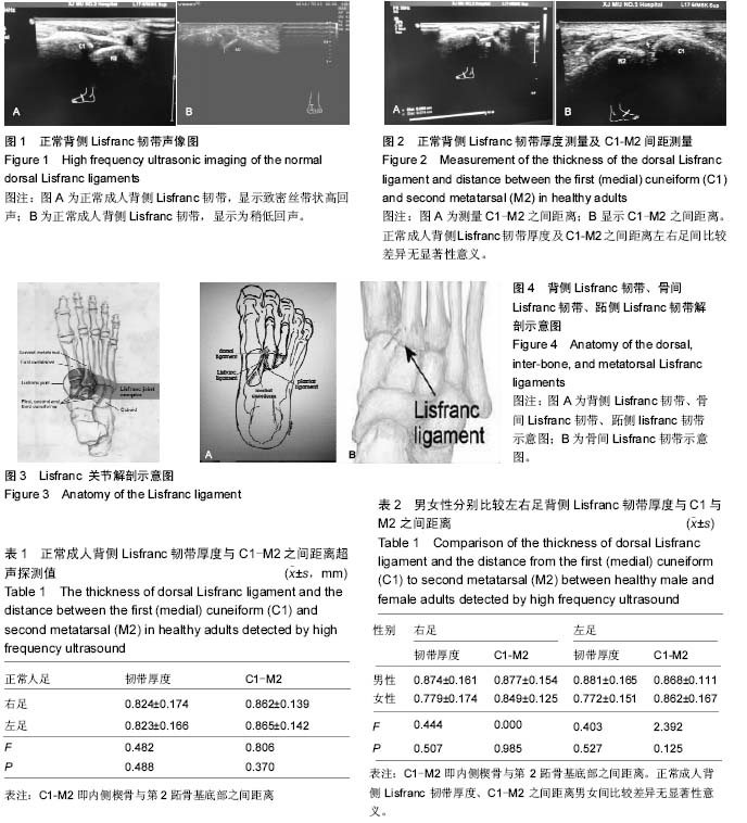
结果与结论:①高频超声对100例志愿者(200个)背侧Lisfranc韧带的显示率为100%;②正常成人背侧Lisfranc韧带厚度右足与左足比较差异无显著性意义(P > 0.05);③男性背侧Lisfranc韧带右足、左足厚度与女性背侧Lisfranc韧带右足、左足厚度比较差异无显著性意义(P > 0.05);④C1与M2之间距离右足与左足比较差异无显著性意义(P > 0.05),C1与M2之间距离男性右足、左足与女性右足、左足比较差异无显著性意义(P > 0.05);⑤结果说明,高频超声对背侧Lisfranc韧带具有良好的显示效果;测量C1与M2之间的距离也较方便、准确,是一种可行、无创、价廉的检查方法,可作为Lisfranc关节损伤早期诊断的间接征象指标的影像学检查方法。
中国组织工程研究杂志出版内容重点:组织构建;骨细胞;软骨细胞;细胞培养;成纤维细胞;血管内皮细胞;骨质疏松;组织工程
ORCID: 0000-0003-3448-5942(陈佩瑾)
中图分类号:
.jpg) 文题释义:
Lisfranc损伤:Lisfranc关节即通常所指的跖跗关节,1815年法国外科医师Jaqcues Lisfranc de Saint- Martin在为一位士兵治疗前足坏疽截肢时,发现通过这个关节可以不用截断骨骼,且省时省力,可取得较好的效果,从此以其名字命名。Lisfranc损伤的狭义定义仅指发生在跖跗关节部位的损伤,其所包括的范围涉及跖跗关节的骨性或韧带组织的损伤,而广义的定义不仅包括跖跗关节,还包括近侧跖骨间关节以及跗骨间关节的骨性或韧带组织的损伤。
Lisfranc韧带:Lisfranc韧带起自内侧楔骨的外侧缘,向远端、跖外侧方向斜形走行,止于第2跖骨基底部内侧近关节面处。第2-5跖骨基底之间有横向的韧带连接,而第1,2跖骨基底之间没有横向的韧带,所以Lisfranc韧带是唯一连接于第1,2跖骨之间的韧带连接,其完整性对于关节的稳定十分重要。Lisfranc 韧带是跖跗关节复合体中最坚强的。它将第2跖骨基底部 “锁定”在原位,以保证基石样结构的稳定性。
文题释义:
Lisfranc损伤:Lisfranc关节即通常所指的跖跗关节,1815年法国外科医师Jaqcues Lisfranc de Saint- Martin在为一位士兵治疗前足坏疽截肢时,发现通过这个关节可以不用截断骨骼,且省时省力,可取得较好的效果,从此以其名字命名。Lisfranc损伤的狭义定义仅指发生在跖跗关节部位的损伤,其所包括的范围涉及跖跗关节的骨性或韧带组织的损伤,而广义的定义不仅包括跖跗关节,还包括近侧跖骨间关节以及跗骨间关节的骨性或韧带组织的损伤。
Lisfranc韧带:Lisfranc韧带起自内侧楔骨的外侧缘,向远端、跖外侧方向斜形走行,止于第2跖骨基底部内侧近关节面处。第2-5跖骨基底之间有横向的韧带连接,而第1,2跖骨基底之间没有横向的韧带,所以Lisfranc韧带是唯一连接于第1,2跖骨之间的韧带连接,其完整性对于关节的稳定十分重要。Lisfranc 韧带是跖跗关节复合体中最坚强的。它将第2跖骨基底部 “锁定”在原位,以保证基石样结构的稳定性。