| [1] Takahashi K, Yamanaka S. Induction of pluripotent stem cells from mouse embryonic and adult fibroblast cultures by defined factors. Cell. 2006;126(4): 663-676.[2] Takahashi K, Tanabe K, Ohnuki M, et al. Induction of pluripotent stem cells from adult human fibroblasts by defined factors. Cell. 2007;131(5): 861-872.[3] Yu J, Vodyanik MA, Smuga-Otto K, et al. Induced pluripotent stem cell lines derived from human somatic cells. Science. 2007;318(5858): 1917-1920.[4] Cheng F, Ke Q, Chen F, et al. Protecting against wayward human induced pluripotent stem cells with a suicide gene. Biomaterials. 2012;33(11): 3195-3204.[5] 张杨,李秀兰.诱导性多潜能干细胞的安全性及临床应用[J].中国组织工程研究, 2013,17(27):5087-5093.[6] Li W, Xiang AP. Safeguarding clinical translation of pluripotent stem cells with suicide genes. Organogenesis. 2013;9(1):34-39.[7] 姚欣鹏,徐旸,张璐,等.分子影像学方法示踪胚胎干细胞移植治疗急性肝损伤[J].中国组织工程研究,2013,17(36): 6481-6488.[8] Landy A. Dynamic, structural, and regulatory aspects of lambda site-specific recombination. Annu Rev Biochem. 1989;58: 913-949.[9] Furuya M, Yasuchika K, Mizutani K, et al. Electroporation of cynomolgus monkey embryonic stem cells. Genesis. 2003;37(4): 180-187.[10] Ueda S, Yoshikawa M, Ouji Y, et al. Cynomolgus monkey embryonic stem cell lines express green fluorescent protein. J Biosci Bioeng. 2006;102(1): 14-20.[11] Suter DM, Cartier L, Bettiol E, et al. Rapid generation of stable transgenic embryonic stem cell lines using modular lentivectors. Stem Cells. 2006;24(3): 615-623.[12] Belfield EJ, Hughes RK, Tsesmetzis N, et al. The gateway pDEST17 expression vector encodes a -1 ribosomal frameshifting sequence. Nucleic Acids Res. 2007;35(4): 1322-1332.[13] Chen QJ, Zhou HM, Chen J, et al. A Gateway-based platform for multigene plant transformation. Plant Mol Biol. 2006;62(6): 927-936.[14] Sivagnanam S, Majumdar A, Yoshimoto K, et al. Early experiences in developing and managing the neuroscience gateway. Concurr Comput. 2015;27(2): 473-488.[15] Wu TM, Lin KC, Liau WS, et al. A set of GFP-based organelle marker lines combined with DsRed-based gateway vectors for subcellular localization study in rice (Oryza sativa L.). Plant Mol Biol. 2016;90(1-2): 107-115. [16] Griffin TZ, Kang W, Ma Y, et al. The HAND Database: a gateway to understanding the role of HIV in HIV-associated neurocognitive disorders. BMC Med Genomics. 2015;8(1):70.[17] Silva J, Nichols J, Theunissen TW, et al. Nanog is the gateway to the pluripotent ground state. Cell, 2009; 138(4): 722-737.[18] Galvagni F, Neri F.Snail represses Nanog to promote embryonic stem cell differentiation. Genom Data. 2015; 4:82-83.[19] 沈洁,李金辉,阮静,等.诱导多能干细胞建系及体外造血祖细胞定向分化体系的建立[J].中国组织工程研究,2014, 18(19): 3005-3011.[20] Evans M. Discovering pluripotency: 30 years of mouse embryonic stem cells. Nat Rev Mol Cell Biol. 2011; 12(10): 680-686.[21] Kiuru M, Boyer JL, O'Connor TP, et al. Genetic control of wayward pluripotent stem cells and their progeny after transplantation. Cell Stem Cell. 2009;4(4): 289-300.[22] Wilkinson AC, Goode DK, Cheng YH, et al. Single site-specific integration targeting coupled with embryonic stem cell differentiation provides a high-throughput alternative to in vivo enhancer analyses. Biol Open. 2013;2(11):1229-1238.[23] Xia X, Zhang Y, Zieth CR, et al. Transgenes delivered by lentiviral vector are suppressed in human embryonic stem cells in a promoter-dependent manner. Stem Cells Dev. 2007;16(1): 167-176.[24] 蔡弢艺,陈雄生,贾连顺,等.碱性成纤维细胞生长因子重组腺病毒转染兔骨髓间充质干细胞的表型变化[J].中国组织工程研究,2014,18(23):3727-3731.[25] Smith-Arica JR, Thomson AJ, Ansell R, et al. Infection efficiency of human and mouse embryonic stem cells using adenoviral and adeno-associated viral vectors. Cloning Stem Cells. 2003;5(1): 51-62.[26] Prouty AM, Fischer J, Purdom A, et al. Spiritual Coping: A Gateway to Enhancing Family Communication During Cancer Treatment. J Relig Health. 2016;55(1): 269-287.[27] Amaya CN, Bryan BA. Enrichment of the embryonic stem cell reprogramming factors Oct4, Nanog, Myc, and Sox2 in benign and malignant vascular tumors. BMC Clin Pathol. 2015;15:18.[28] Kang J, Shakya A, Tantin D. Stem cells, stress, metabolism and cancer: a drama in two Octs. Trends Biochem Sci. 2009;34(10):491-499. [29] Liedtke S, Stephan M, Kögler G. et al. Oct4 expression revisited: potential pitfalls for data misinterpretation in stem cell research. Biol Chem. 2008;389(7): 845-850.[30] Tsai LL, Hu FW, Lee SS, et al. Oct4 mediates tumor initiating properties in oral squamous cell carcinomas through the regulation of epithelial-mesenchymal transition. PLoS One. 2014;9(1):e87207. [31] Liu J, Wang L, Ma L, et al. Significantly increased expression of OCT4 and ABCG2 in spheroid body-forming cells of the human gastric cancer MKN-45 cell line. Oncol Lett. 2013;6(4):891-896.[32] Silva J, Nichols J, Theunissen TW, et al. Nanog is the gateway to the pluripotent ground state. Cell. 2009; 138(4): 722-737.[33] Li WZ, Ai ZY, Wang ZW, et al. GATA-1 directly regulates Nanog in mouse embryonic stem cells. Biochem Biophys Res Commun. 2015;465(3):575-579.[34] Guo C, Xue Y, Yang G, et al. Nanog RNA binding proteins YBX1 and ILF3 affect pluripotency of embryonic stem cells. Cell Biol Int. 2015. [Epub ahead of print] |
.jpg)

.jpg)
.jpg)
.jpg) 文题释义:
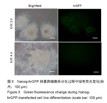
Nanog基因:特定性高表达于多能干细胞,例如诱导多能干细胞与胚胎干细胞,并且随多能干细胞的分化,表达量迅速下调。因此Nanog基因可作胚胎干细胞的分化研究中重要的观察对象,把带有Nanog介导hrGFP载体转入建立纯化的胚胎干细胞系,就可以实时观察胚胎干细胞的干性特征,并可通过观察其分化过程中绿色荧光的表达实时跟踪记录分化过程,这在多能干细胞的分化及分化调控研究中具有十分重要的意义。
Gateway技术:是利用λ噬菌体特异性结合位点,可在Clonase的作用下整合到宿主E.coli的基因组中的一种新的通用型克隆技术。利用Gateway技术构建载体,只需要在目的基因两端加上特异性的 15 bp大小的att结合位点,通过两种Clonase作用下的BP和LR结合反应,即可将目的基因导入载体中,其整合率可达到100%。而且去除了酶切、连接过程,可以避免假阳性的出现。因其操作方便,Gateway技术近年来在基因工程中的应用已越来越广泛。
文题释义:
Nanog基因:特定性高表达于多能干细胞,例如诱导多能干细胞与胚胎干细胞,并且随多能干细胞的分化,表达量迅速下调。因此Nanog基因可作胚胎干细胞的分化研究中重要的观察对象,把带有Nanog介导hrGFP载体转入建立纯化的胚胎干细胞系,就可以实时观察胚胎干细胞的干性特征,并可通过观察其分化过程中绿色荧光的表达实时跟踪记录分化过程,这在多能干细胞的分化及分化调控研究中具有十分重要的意义。
Gateway技术:是利用λ噬菌体特异性结合位点,可在Clonase的作用下整合到宿主E.coli的基因组中的一种新的通用型克隆技术。利用Gateway技术构建载体,只需要在目的基因两端加上特异性的 15 bp大小的att结合位点,通过两种Clonase作用下的BP和LR结合反应,即可将目的基因导入载体中,其整合率可达到100%。而且去除了酶切、连接过程,可以避免假阳性的出现。因其操作方便,Gateway技术近年来在基因工程中的应用已越来越广泛。